Introduction
Students would often wonder, what are the main organs of the respiratory system? What is the function of the respiratory system? What is the main function of the lung in human body?
This article focuses on answering those questions step by step with a brief knowledge of the anatomy and physiology of the human respiratory system.
What are the main organs of the respiratory system? – Anatomy of the respiratory system
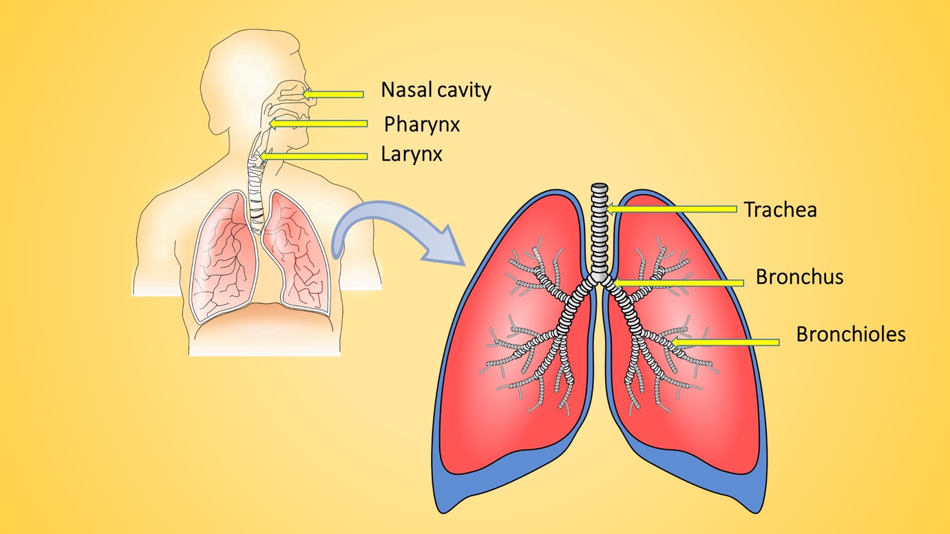
The main parts of the respiratory system are;
- Nose, Mouth, and nasal cavity
- Pharynx (throat)
- Larynx (the voice-box)
- Trachea (the windpipe)
- Lungs
- Bronchi and bronchioles (the airways)
- Alveoli
They are shown in the following diagram of the respiratory system.
What is the main function of the human respiratory system?
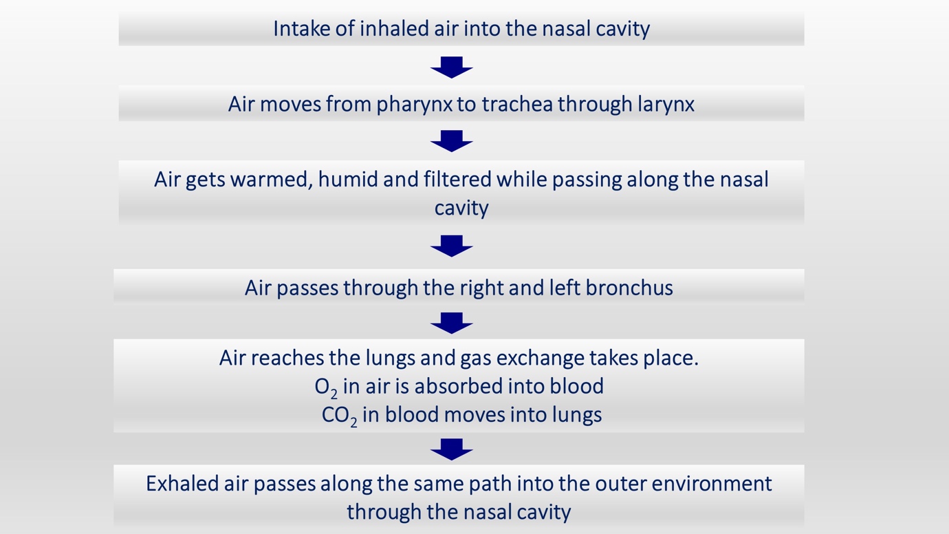
The human respiratory system is involved in transporting oxygen (O2) into the lungs and transporting gaseous waste like carbon dioxide (CO2) out of the human body.
This process is known as the gas exchange in the lungs. Each and every organ of the human respiratory system is adapted to carry out its specific function in this complex process. Let’s see these in detail.
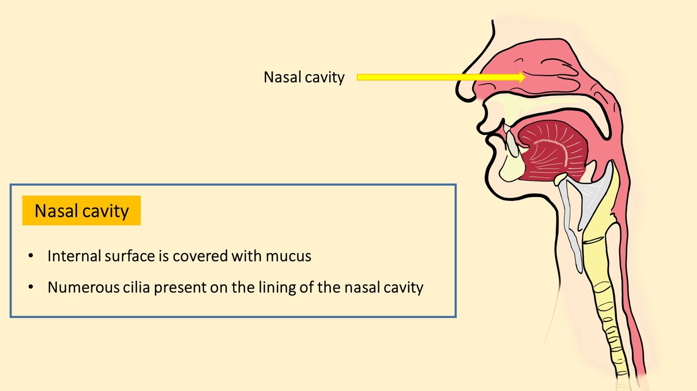
What is the function of the nasal cavity in the respiratory system?
The human nasal cavity acts as the passage for inhaled air. It has a most internal surface and there are cilia in the lining of the nasal cavity.
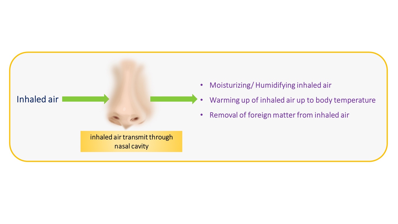
What happens to the air which enters the nose?
Inhaled air undergoes a series of changes for optimum respiration. Air is humidified and warmed to match the internal body temperature. Hair in the nose and the cilia in the epithelial lining help to trap and remove the foreign dust particles in the inhaled air.
Air from the nasal cavity enters the pharynx.
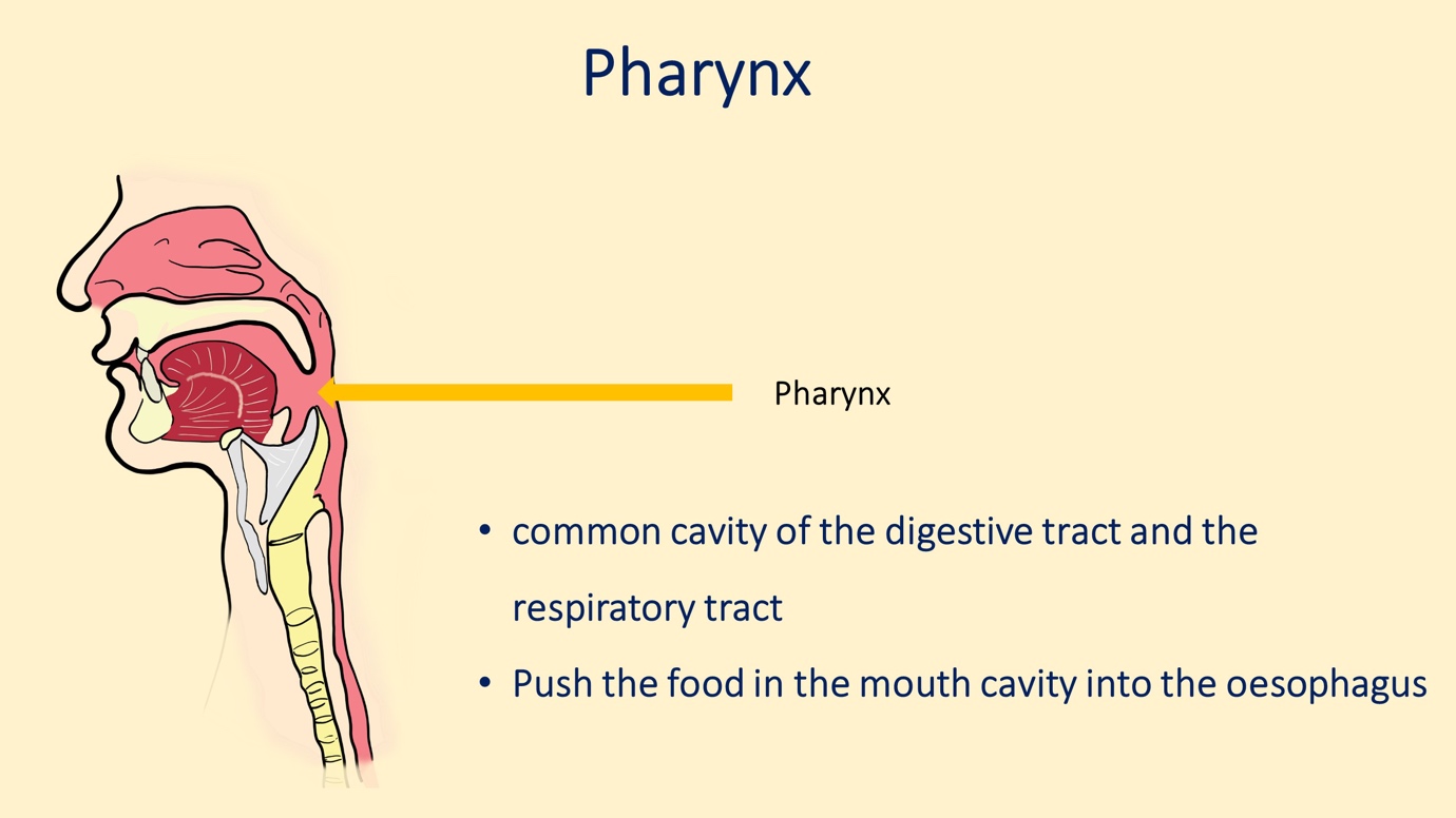
What is the main function of the pharynx?
Students always wonder “ Is the pharynx part of the respiratory system? ” and Is the pharynx part of the digestive system?
Well, the answer to both the questions is YES!
The pharynx acts as a common passage for both respiratory and digestive systems. The pharynx is simply known as “throat”.
Pharynx also humidifies and regulates the temperature of inhaled air and helps in removing foreign particles.
What is the relationship between the larynx, trachea, bronchi, bronchioles, and alveoli?
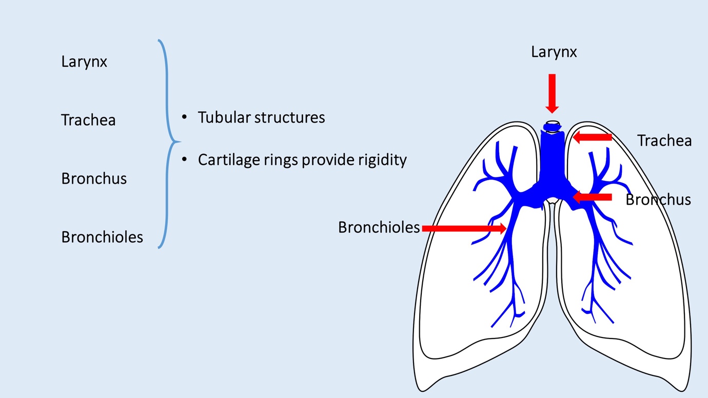
After passing the pharynx air enters into the voice-box, the larynx. Air from the larynx then enters into the trachea (the windpipe). The trachea divides into the left bronchus and the right bronchus. These bronchi enter into lungs.
Inside the lungs, they further divide into bronchioles. Bronchioles are categorized as primary bronchioles, secondary bronchioles, and tertiary bronchioles when they divide into branches.
The larynx, trachea, bronchi, and bronchioles are tubular structures. The larynx, trachea, and bronchi have cartilage rings that prevent collapsing. Bronchioles do not have cartilages. Their patency is maintained by elastic fibers and smooth muscles.
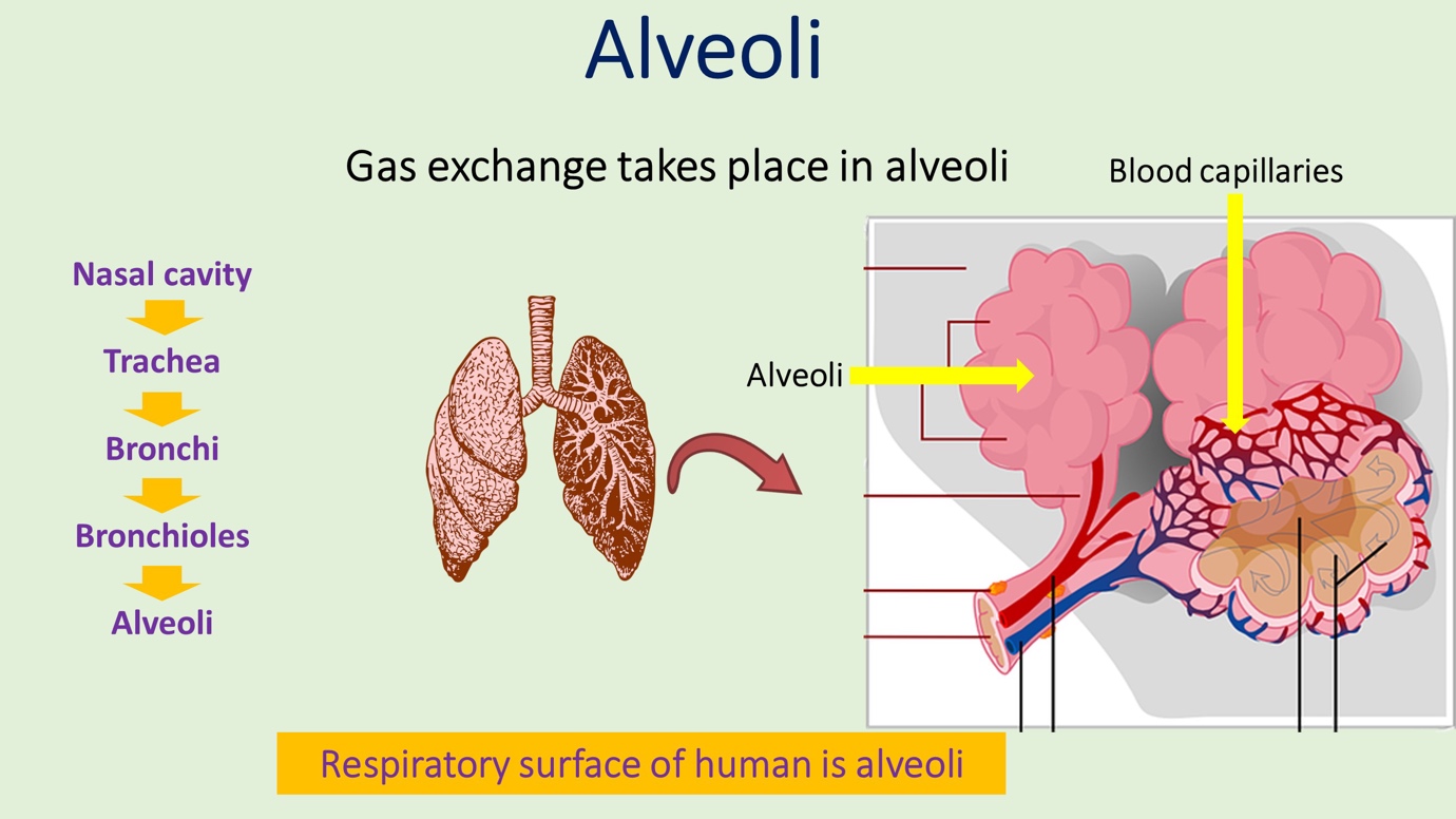
Air from the bronchioles enters into the alveolar air sacs. Bronchioles open into sac-like structures where there is ventilation as well as a proper blood supply. Blood supply is from the fine capillaries in the lung. The special fluid, known as surfactant acts a major role here in maintaining the patency of the alveolar sacs. The gas exchange takes place as alveolar walls. This is described in detail in the next paragraphs.
Lungs
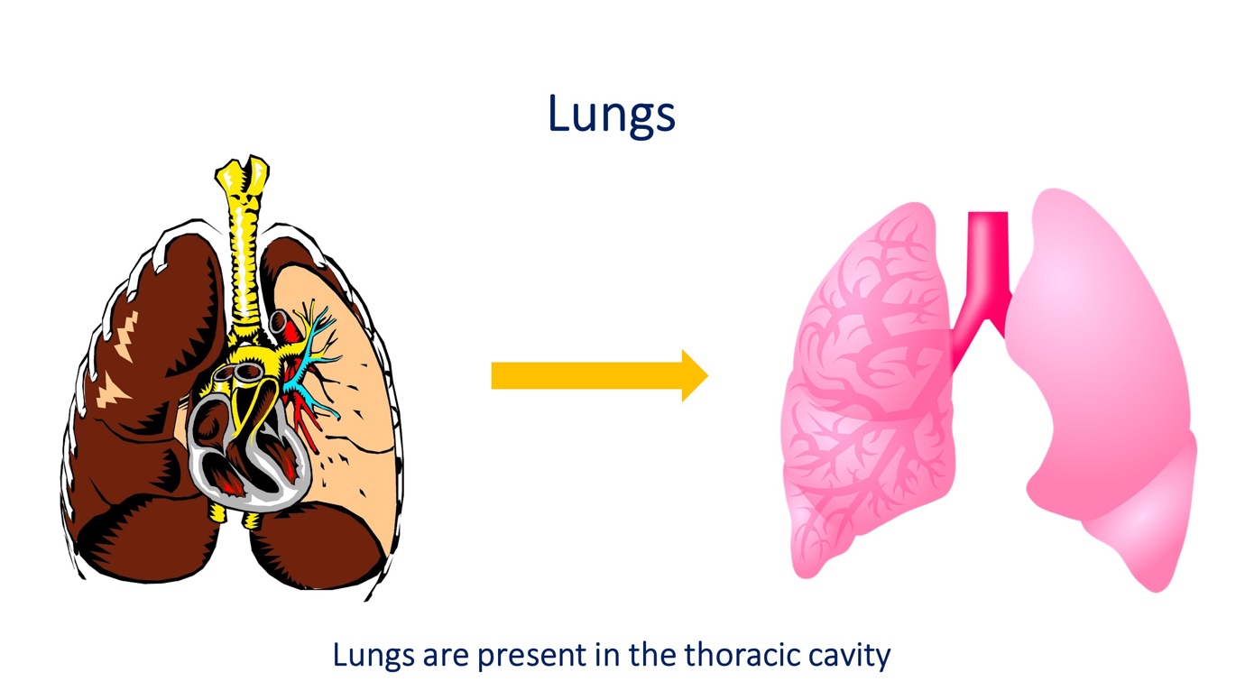
Lungs are spongy structures in the human thoracic cavity. They are a highly vascularized pair of organs. Left lung as an indentation to accommodate the heart. The right lung is made of three lobes and the left lung is made of two lobes.
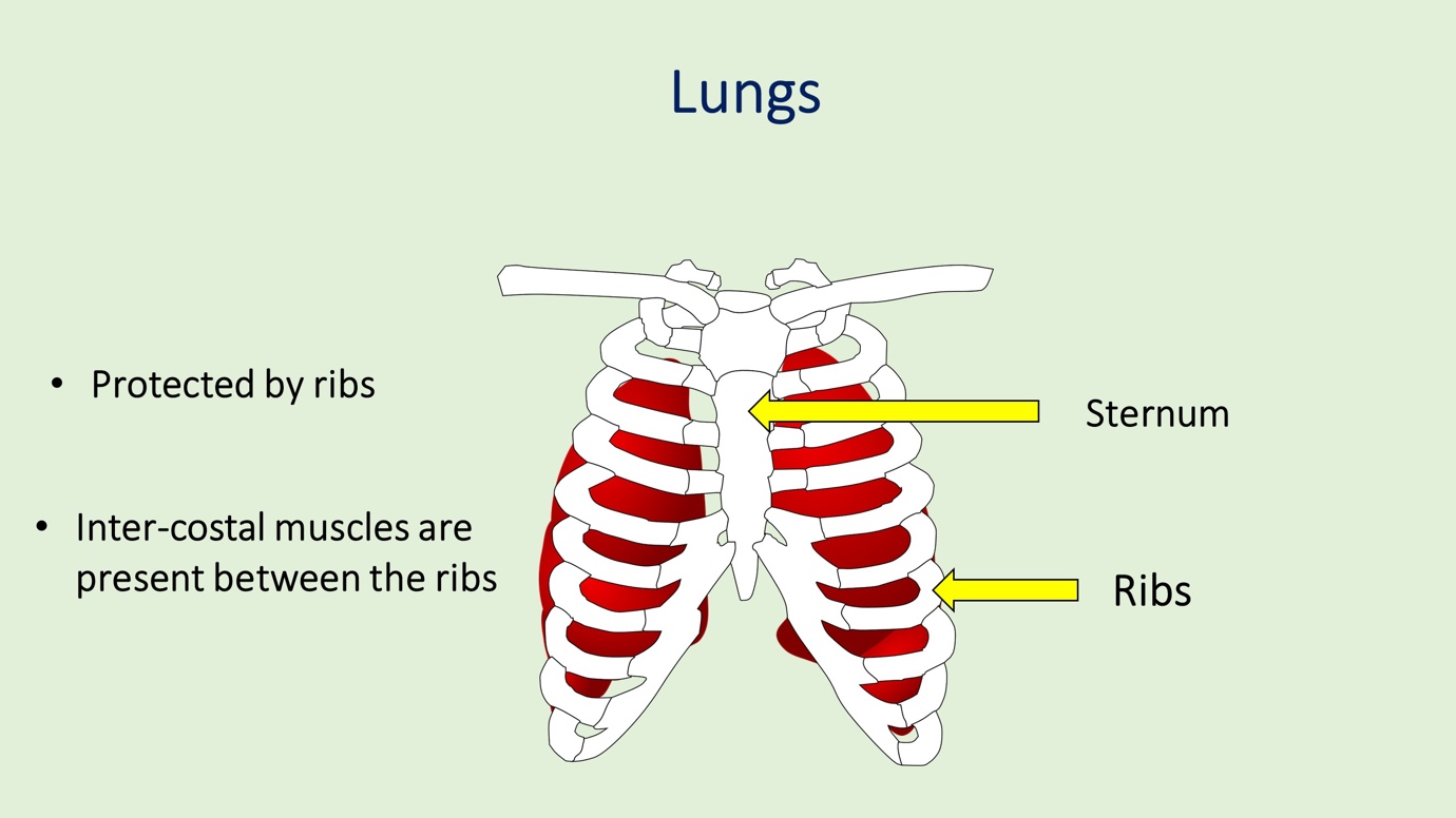
Lungs are protected by the thoracic cage. It is made by the ribs, the sternum, and the intercostal muscles that lie between the ribs. Ribs are anteriorly attached to the sternum and posteriorly attached to the vertebrae.
The lowermost ribs are not attached to the thoracic cage. These free ribs are known as floating ribs.
Physiology of respiratory system simplified – step by step diagram
The function of the respiratory system can be described step by step.
The respiratory surface of humans
The respiratory surface is the place where gas exchange between the external environment and the internal environment (blood) takes place. The respiratory surface of human beings is the wall of alveoli.
At the alveolar level oxygen and carbon, dioxide exchange takes place.
The respiratory surface has specific features for optimum gas exchange. These features are as follows;
- Moist surface
- Permeable for gasses
- Thin surface
- Larger surface area
- Highly vascularized surface
All these features are needed for the proper diffusion of gases across the respiratory surface.
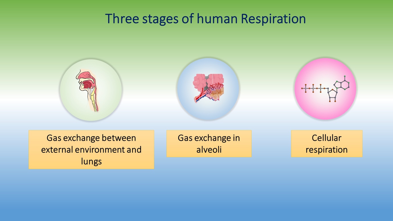
What are the three stages of human respiration?
- Gas exchange with the external environment and the lungs
- Gas exchange in the alveoli
- Cellular respiration
This article focuses on the former two topics. Cellular respiration will be discussed in a separate article.
The first step is carried out by inspiration and expiration. Let’s see these topics in detail.
How do you inhale? What happens when you inhale?
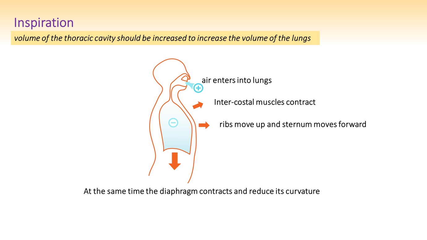
Inspiration, inhalation or simply breathing is the process of taking air into the respiratory system. During inspiration the thoracic cavity and the lung increase in its volume to reduce its pressure. Therefore, inside the lungs and the thoracic cavity, there is a relatively low pressure compared to the outer environment. This pressure gradient causes airflow into the lungs from the external environment.
Increasing the volume of the thoracic cavity is done by the following mechanisms;
- Inter-costal muscles contract – These are the muscles that lie between the ribs
- Ribs move up
- Sternum moves forward
- Diaphragm contracts – its curvature is reduced
These muscle movements altogether expand the thoracic cavity in inhalation.
How do you exhale? What happens when you exhale?
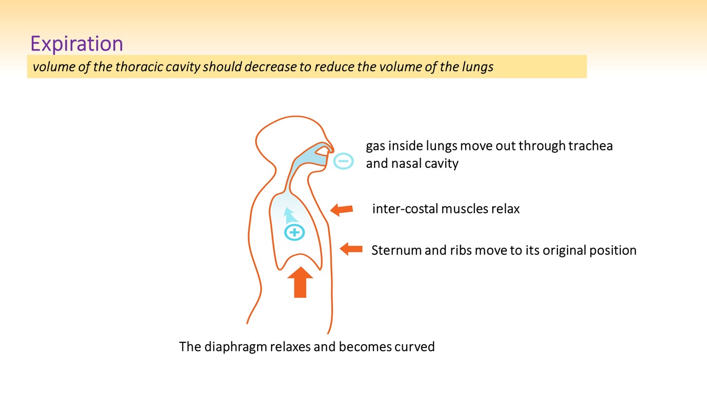
Expiration, exhalation or simply breathing out is the process of removing gaseous waste from the respiratory system. For the process of expiration, the pressure gradient of gases should be from the lungs towards the external environment. That means the pressure inside the lungs and the thoracic cavity has to be increased. This is done by reducing the volume of the lungs and thoracic cavity.
Reducing the volume of the thoracic cavity is done by the following mechanisms;
- Inter-costal muscles relax
- Ribs move towards the original position
- Sternum moves towards the previous position
- The diaphragm relaxes – its curvature is increased
These muscle movements altogether contract the thoracic cavity in exhalation.
Gas exchange in the respiratory surface
Gas exchanges take place at alveoli. Alveoli altogether contribute to a massive surface area.
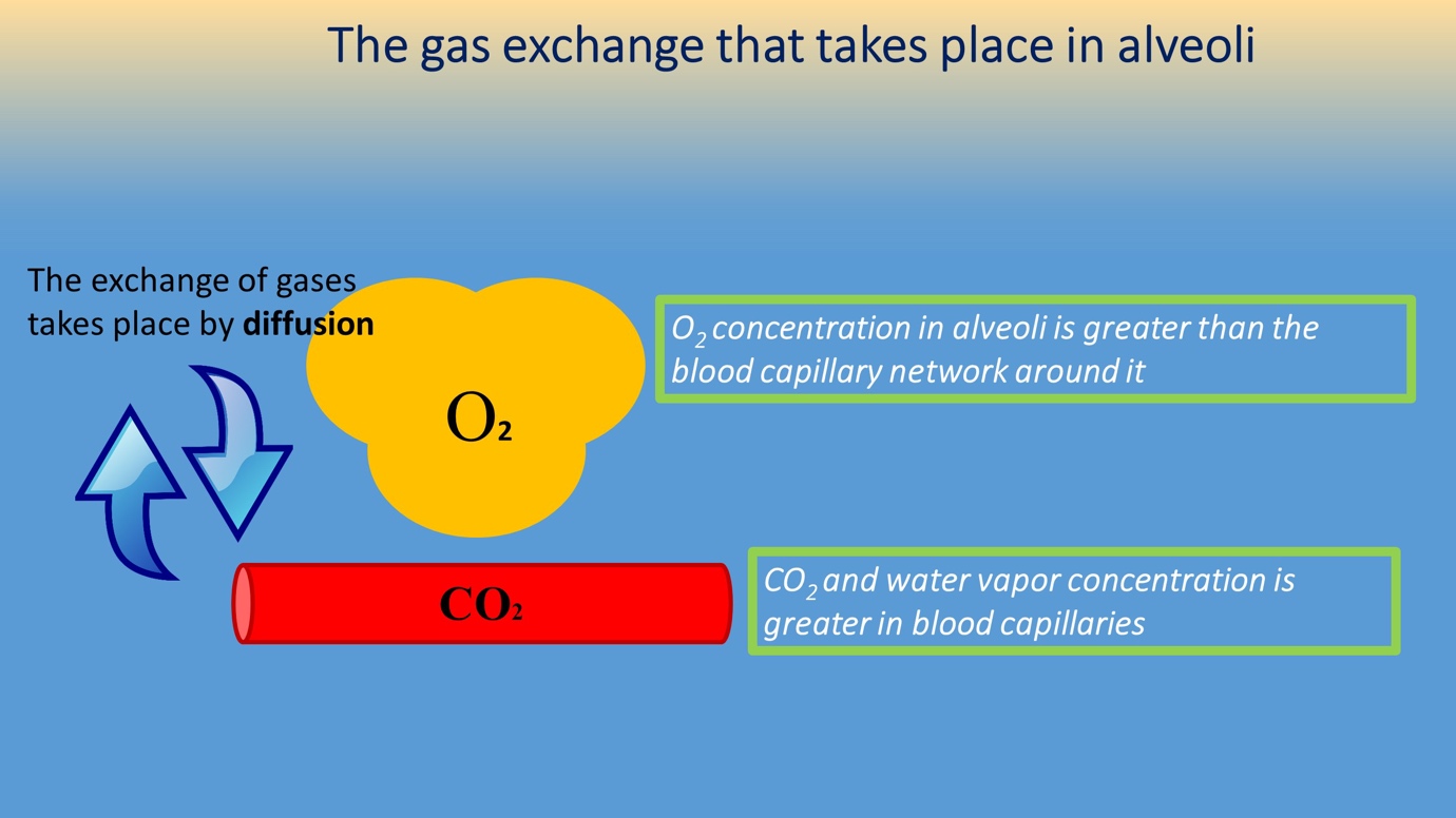
Gas exchange is based on diffusion. In diffusion, particles flow along a concentration gradient without using energy. That means particles from the higher concentration area flows towards the lower concentration area. This principle is also seen in the gas exchange.
Alveoli are filled with inhaled air and blood capillaries are filled with deoxygenated blood and gaseous waste like carbon dioxide. So, the oxygen concentration in alveoli is high and the oxygen concentration in the blood capillaries is low. Because of diffusion, oxygen flows along a concentration gradient. Therefore, oxygen from alveoli enters the blood capillaries.
Let’s consider the concentration of carbon dioxide in alveoli and blood capillaries. Inside blood capillaries, there is a low amount of oxygen and a high amount of carbon dioxide (deoxygenated blood). Carbon dioxide concentration in the blood capillaries is high. Carbon dioxide concentration in the alveoli is low. So, carbon dioxide flows along a concentration gradient according to diffusion. Therefore, the flow of carbon dioxide is from the blood capillaries to the alveoli.
