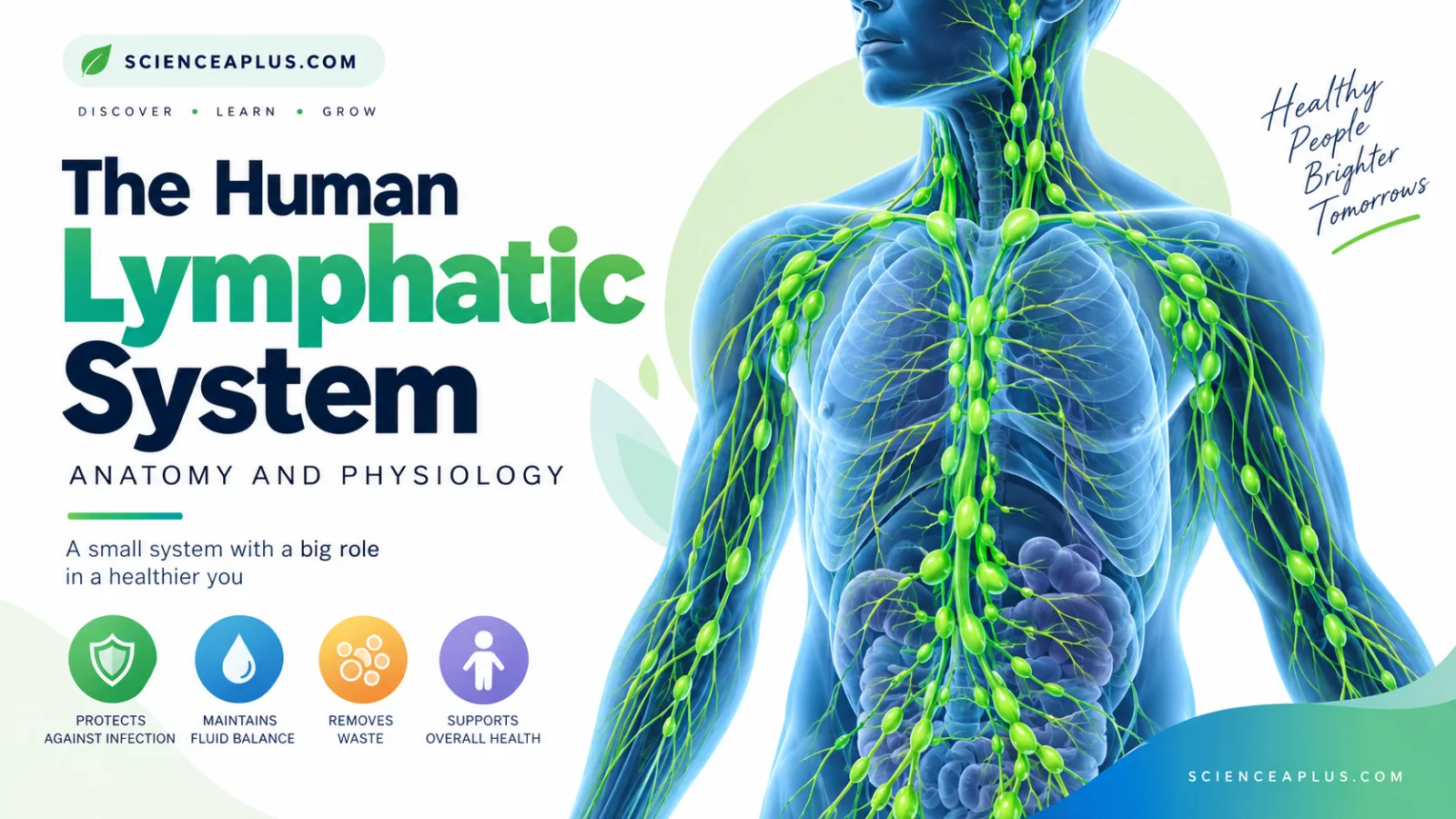
Every day, about 20 litres of fluid leak out of your blood capillaries, and in the standard textbook estimate around 3 litres of it gets back by a separate route. The lymphatic system is the network of vessels, lymph nodes and lymphoid organs that collects this fluid, filters it for germs and returns it to the blood, while also absorbing fats from your gut and housing the white blood cells that defend you.
This guide explains the human lymphatic system from the ground up: what lymph is, how it forms, how it differs from blood, and why your body could not work without it. If you are revising for GCSE biology, the diagrams, flip cards and exam questions later in the article are written for you.
What is the lymphatic system?
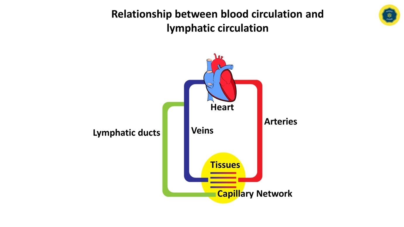
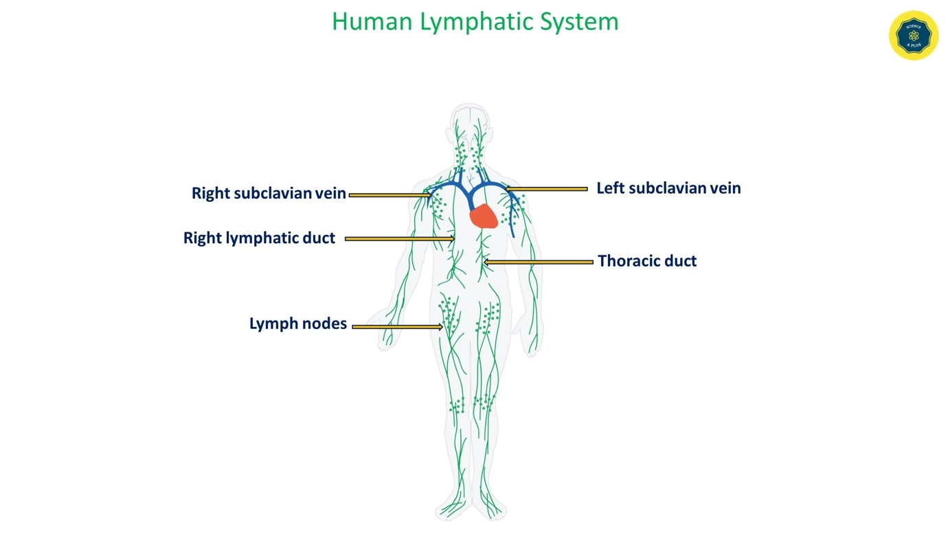
The human lymphatic system is a network of vessels, lymph nodes and lymphoid organs and tissues that runs alongside your blood circulation. Unlike the blood system, it is not a loop. It starts as tiny blind-ended tubes in your tissues and ends by emptying into large veins near the base of the neck.
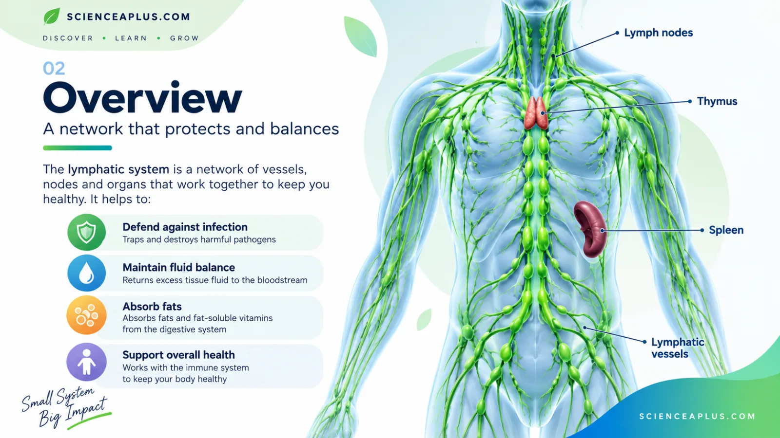
The fluid inside these vessels is called lymph. It is clear to pale yellow and contains water, ions, some proteins and lymphocytes, which are a type of white blood cell. You can think of the lymphatic system as a drainage and security network that works quietly beside the blood circulation. If you want the bigger picture first, our guide to blood and circulation in animals sets the scene.
The system is made of three connected parts: the vessels that carry lymph, the lymph nodes that filter it, and the lymphoid organs and tissues (such as the thymus, spleen and tonsils) where lymphocytes are produced, matured and stored.
The three main jobs
The functions of the lymphatic system fall into three groups. Each one gets its own section later in this article.
- Fluid balance: returning excess tissue fluid to the blood so that tissues do not swell.
- Fat absorption: absorbing fats and fat-soluble vitamins from the small intestine through vessels called lacteals.
- Immune defence: lymph nodes filter lymph, and lymphocytes (B cells and T cells) are produced, matured and stored in lymphoid organs. For the wider story of how your body fights infection, see our introduction to the human immune system.
How tissue fluid and lymph form
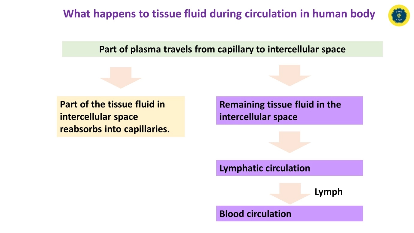
Lymph does not appear from nowhere. It begins life as tissue fluid, the watery liquid that bathes every cell in your body. To understand lymph, you first need to see how tissue fluid forms and where it goes.
At the arterial end
Blood enters a capillary bed from an artery at high pressure. This high hydrostatic (blood) pressure pushes water and small dissolved substances out through the thin capillary walls. These include glucose, amino acids, ions and oxygen. Large plasma proteins and red blood cells are too big to leave, so they stay in the blood.
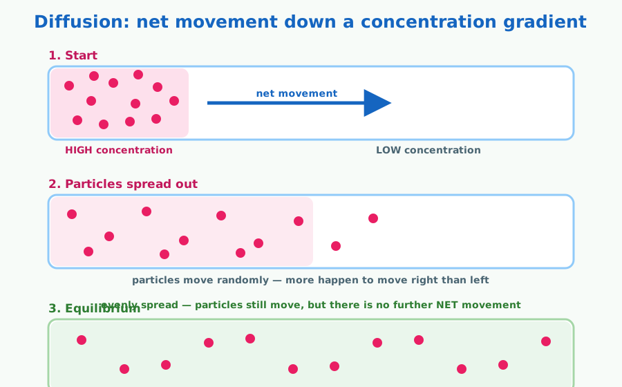
The fluid that escapes is tissue fluid. It delivers oxygen and nutrients to the cells and collects their waste. The substances move between blood and cells partly by diffusion, which is why the capillary walls are so thin.
At the venous end
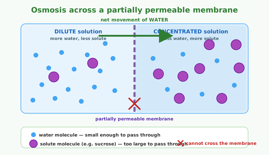
By the time blood reaches the far end of the capillary, its hydrostatic pressure has dropped. Meanwhile the plasma proteins left behind give the blood a low water potential (this is the oncotic pressure). With less pushing out and more pulling in, most of the tissue fluid is drawn back into the capillary by osmosis.
What happens to the leftover fluid
Not all of the fluid returns to the capillaries. Roughly 85 % does (about 17 of the 20 litres), but about 15 % drains into lymph capillaries instead. Once it has entered those vessels, it is called lymph.
In an adult, about 20 litres of fluid filter out of the capillaries each day. Around 17 litres are reabsorbed, and the remaining 3 litres return through the lymphatic system. Without that return route, fluid would build up in the tissues and the blood volume would fall. The lymph eventually rejoins the blood, completing a one-way trip that complements the double blood circulation of the heart and lungs.
20 L filtered → 17 L reabsorbed → 3 L returned as lymph
Lymph vs blood vs tissue fluid
Students often mix up these three fluids, and exam questions love to test the differences. All three are related: tissue fluid comes from blood plasma, and lymph is tissue fluid that has entered the lymph capillaries. The table below puts them side by side. For more on what blood itself contains, read our complete lesson on human blood.
| Feature | Blood | Tissue fluid | Lymph |
|---|---|---|---|
| Where it is found | Inside closed blood vessels | Bathing the cells in the tissues | Inside lymph vessels |
| What moves it | The pumping heart | Pressure differences across the capillary wall | Muscle squeezing, breathing movements, vessel walls and valves |
| Red blood cells | Yes | No | No |
| Lymphocytes | Yes (one type of white blood cell) | Few | Yes, many |
| Platelets | Yes | No | No |
| Plasma proteins | Lots | Little | Little |
| Fats | Carried in blood after absorption | Not a main feature | Present as chylomicrons after meals (milky chyle from the gut) |
The key pattern to remember is that blood is pumped and protein-rich, tissue fluid has no red cells and little protein, and lymph is moved by muscles and valves and carries lymphocytes.
Lymphatic vessels and ducts
The lymphatic system has its own set of vessels, running alongside the blood vessels in almost every tissue. Unlike the blood system, it is not a loop. Lymph flows in one direction only, from the tissues towards the neck, where it rejoins the blood.
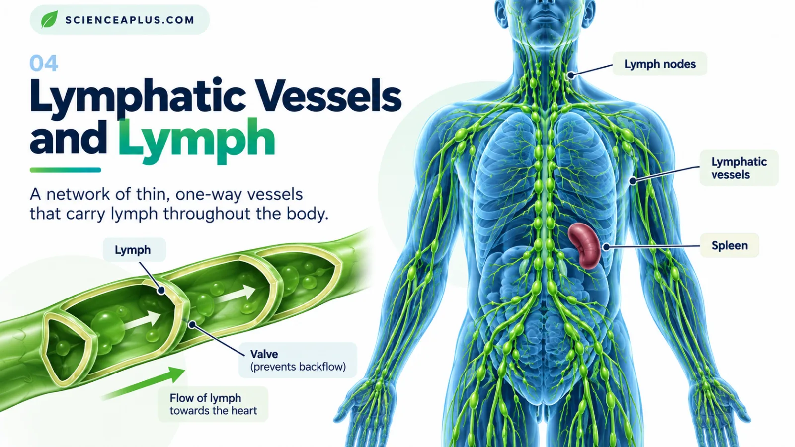
The vessels get steadily wider as they travel. Tiny lymph capillaries join to form larger lymph vessels, and these in turn join two main ducts that empty into veins near the base of the neck.
Lymph capillaries
Lymph capillaries are where tissue fluid first enters the lymphatic system. They are blind-ended, which means they are closed at one end, rather than forming a loop like blood capillaries.
Their walls are made of overlapping endothelial cells. These act like one-way flaps: when pressure in the surrounding tissue fluid rises, the flaps are pushed open and fluid flows in. When pressure inside the capillary is higher, the flaps close, so lymph cannot leak back out.
Lymph capillaries are also more permeable than blood capillaries. That matters because they can take up large molecules, including proteins, that are too big to go back into blood capillaries easily.
Valves and how lymph moves (no central pump)
Here is a question students often ask: does the lymphatic system have a pump? Not a central one. The blood has the heart, but lymph has no single organ that pumps it. If you want to compare it with the pumped system, see our guide to the cardiac cycle and blood circulation.
Instead, lymph is moved along by several everyday forces:
- Skeletal muscles squeeze the lymph vessels as you move, pushing lymph onwards.
- Breathing changes the pressure in the chest, which helps draw lymph upwards.
- Smooth muscle in the walls of the larger lymph vessels contracts and moves lymph along.
- Valves inside the larger vessels, much like those in veins, stop lymph flowing backwards.
The thoracic duct and right lymphatic duct
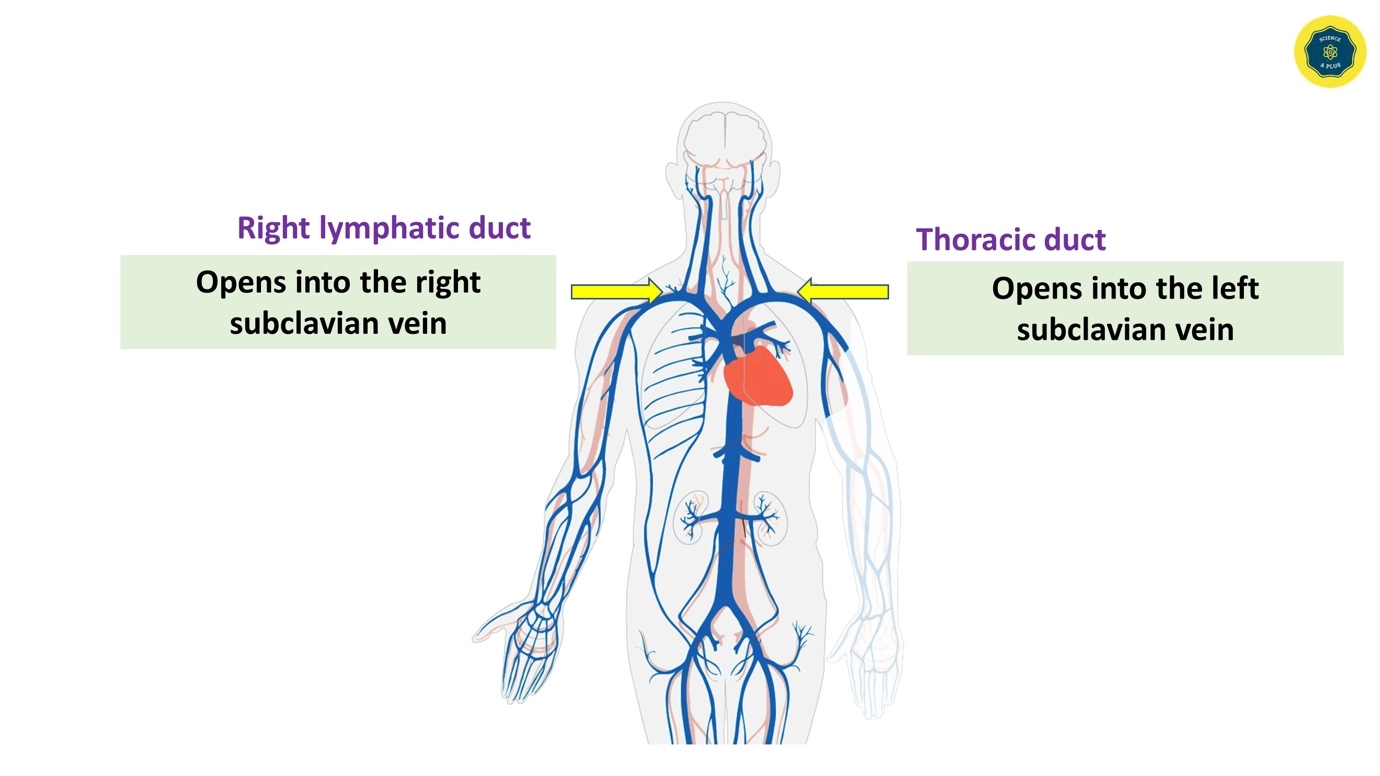
All the lymph in the body eventually drains into one of two ducts, and each one empties into the blood at a junction of two veins.
The thoracic duct is the largest lymph vessel in the body. It drains lymph from the legs, the abdomen, the left arm, and the left side of the head, neck and chest. It empties into the left venous angle, where the left subclavian vein and the left internal jugular vein meet.
The right lymphatic duct is much smaller. It drains the right arm and the right side of the head, neck and chest into the junction of the right subclavian vein and the right internal jugular vein.
At the start of the thoracic duct is a sac called the cisterna chyli. It collects lymph from the abdomen and legs before the duct carries it upwards.
| Duct | Drains lymph from | Empties into |
|---|---|---|
| Thoracic duct | Legs, abdomen, left arm, left side of head, neck and chest | Left venous angle (left subclavian and left internal jugular veins) |
| Right lymphatic duct | Right arm, right side of head, neck and chest | Junction of right subclavian and right internal jugular veins |
Trace the path of lymph
The best way to understand the human lymphatic system is to follow a drop of fluid from start to finish. Here is the journey, step by step.
- Blood capillary (arterial end): high hydrostatic pressure pushes water and small solutes such as glucose, amino acids, ions and oxygen out through the capillary wall. Large plasma proteins and red blood cells stay inside.
- Tissue fluid: the fluid that has escaped bathes the surrounding cells, delivering nutrients and collecting waste.
- Venous end: the hydrostatic pressure is lower and plasma proteins pull water back by osmosis, so most of the fluid returns to the blood capillaries.
- Lymph capillary: the leftover fluid, roughly 15 % of the total, enters a blind-ended lymph capillary through the one-way flaps. It is now called lymph.
- Lymph vessels: lymph is moved along by muscle movement, breathing and smooth muscle, with valves preventing backflow.
- Lymph nodes: on the way, lymph passes through lymph nodes, where it is filtered and checked for pathogens.
- Duct to vein: lymph reaches the thoracic duct or the right lymphatic duct, and then flows into a vein at the base of the neck, rejoining the blood.
Over a day, about 20 litres of fluid filter out of the capillaries of an adult. Around 17 litres are reabsorbed directly, and about 3 litres return through the lymphatic system.
Exam question: describe how tissue fluid returns to the blood. (4 marks)
At the venous end of the capillary, the hydrostatic pressure is lower than at the arterial end. Plasma proteins remain in the blood and give it a low water potential, so water moves back into the capillary by osmosis. This returns most of the tissue fluid, about 85 %. The remaining fluid, about 15 %, enters the blind-ended lymph capillaries and becomes lymph. Lymph flows through lymph vessels and ducts and empties into veins near the neck, so it returns to the blood.
Lacteals: how the lymphatic system absorbs fats
Besides draining fluid, the lymphatic system has a second job in digestion. Fats and fat-soluble vitamins are absorbed from the small intestine through special lymph capillaries called lacteals.
Each villus, the tiny finger-like projection lining the small intestine, has a lacteal running through its centre. Most digested nutrients, such as glucose and amino acids, pass into the blood capillaries of the villus. Fats are different.
After digestion, fats are absorbed and packaged into particles called chylomicrons. These are too big to squeeze into blood capillaries, so they enter the lacteals instead. The fluid then travels through lymph vessels to the cisterna chyli and up the thoracic duct, where it finally reaches the bloodstream.
After a meal containing fat, the lymph from the intestine looks milky because of all these chylomicrons. This fat-rich lymph is called chyle. Normal lymph elsewhere in the body is clear to pale yellow.
Lymph nodes
Lymph nodes are small, bean-shaped structures that sit along the lymph vessels of the human lymphatic system. Most are only a few millimetres to about 1–2 cm long. Estimates of how many we have vary from about 450 to 800, so the honest answer is "several hundred".
Think of them as checkpoints. Lymph flows in, is inspected for pathogens, and flows out again cleaner than it arrived.
Where are lymph nodes?
Lymph nodes are scattered throughout the body, but they gather in clusters at key points where lymph from large areas of tissue is collected. The clusters you are most likely to notice are:
- Neck (cervical nodes): drain the head, face and throat.
- Armpits (axillary nodes): drain the arms and the chest wall.
- Groin (inguinal nodes): drain the legs and lower body.
- Chest and abdomen: deeper clusters that you cannot feel from the outside.
Because each cluster serves a particular region, a swollen node can sometimes hint at where an infection is. A sore throat, for example, often makes the neck nodes tender.

What do lymph nodes do?
Each node has several afferent vessels bringing lymph in and one efferent vessel carrying it out. Having more vessels going in than out slows the flow down, which gives the cells inside the node time to do their work.
Inside, two types of cell do the checking. Macrophages trap and destroy pathogens and cancer cells. Lymphocytes (B cells and T cells) recognise particular invaders and mount a targeted immune response. Together they make lymph nodes one of the immune system’s main meeting points.
Why do lymph nodes swell?
During an infection, the lymphocytes inside a node multiply to fight the pathogen. The node fills with extra cells and becomes larger and often tender. These are the "swollen glands" people talk about, such as the neck nodes during a sore throat.
Swollen lymph nodes are usually a sign that your body is fighting an infection, and they normally go down on their own within 1 to 2 weeks. If you are curious about other possible triggers, see our article on whether smoking can cause swollen lymph nodes.
Lymphoid organs
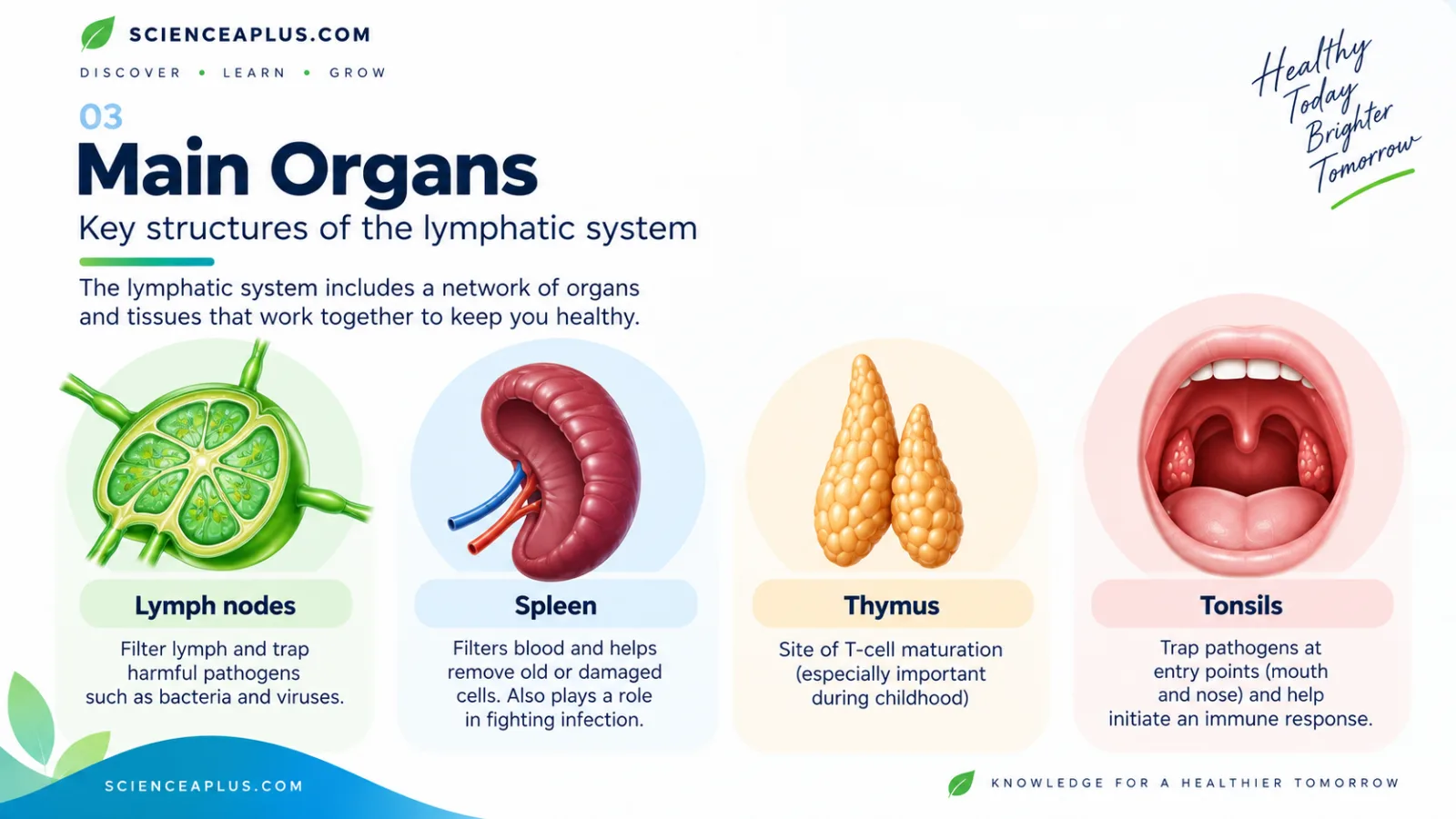
Lymph nodes are only one part of the lymphatic system's immune machinery. Lymphoid organs are the places where lymphocytes are produced, matured and stored. They fall into two groups.
Primary lymphoid organs are where lymphocytes are made and mature: the red bone marrow and the thymus. Secondary lymphoid organs are where mature lymphocytes meet pathogens: lymph nodes, the spleen, the tonsils and adenoids, MALT (such as Peyer's patches in the small intestine) and the appendix.
The spleen
The spleen is a fist-sized organ in the upper left abdomen, tucked under the ribs. It is the largest lymphoid organ, but it filters blood, not lymph. The red pulp removes old and damaged red blood cells and stores some platelets and white cells, while the white pulp contains lymphocytes that respond to pathogens in the blood.
People can live without a spleen, but they have a higher risk of infection. The NHS advises vaccinations and sometimes antibiotics for them. To learn how everyday habits relate to this organ, read about lifestyle choices and a healthy spleen.
The thymus
The thymus is where T cells finish maturing, which is what the "T" stands for. It is large in children, shrinks after puberty and is gradually replaced by fat. By then, most of the body's T cell training has already been done.
Red bone marrow
All blood cells, including lymphocytes, are made in red bone marrow, and B cells mature there. This makes bone marrow the starting point for much of the immune system. The process begins with stem cells, explained in our article on haematopoietic stem cells and differentiation.
Functions of the lymphatic system
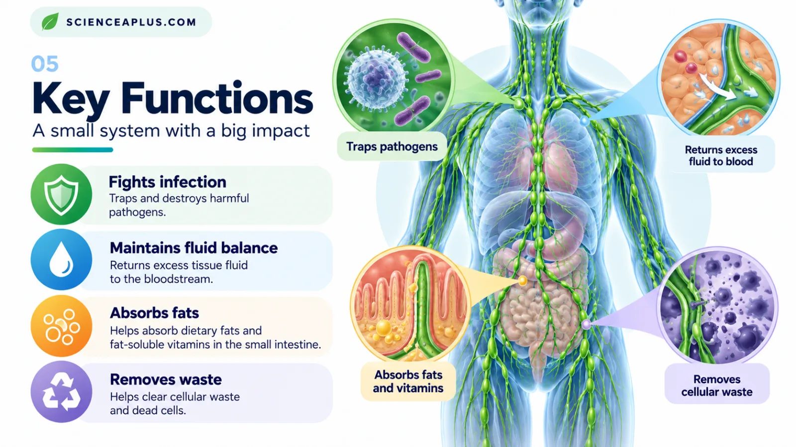
The functions of the lymphatic system fall into a few clear jobs. It keeps the fluid around your cells in balance, carries absorbed fats away from the gut, and gives the immune system places to meet and destroy pathogens. It also helps clear debris from tissues.
1. Fluid balance
Blood capillaries leak fluid all the time. In an adult, about 20 litres filter out each day, roughly 17 litres are reabsorbed by the capillaries, and about 3 litres are left over. Without the lymphatic system to collect that leftover fluid, it would build up in the tissues and cause swelling.
Once the fluid enters lymph capillaries it is called lymph. The lymph travels through vessels and nodes and is returned to the blood near the base of the neck, which keeps blood volume steady.
2. Fat absorption
Fats from your food are absorbed in the small intestine as tiny particles called chylomicrons. These are too big to enter blood capillaries, so they pass into the lacteals in the middle of each villus instead. The fat-rich lymph, called chyle, looks milky and eventually reaches the blood through the thoracic duct. Fat-soluble vitamins travel the same way.
3. Immune defence
Lymph nodes act as filters. Lymph flows in through several afferent vessels and leaves through one efferent vessel, and on the way macrophages and lymphocytes trap and destroy pathogens. Lymphocytes (B cells and T cells) are produced, matured and stored in the lymphoid organs, ready to respond. This is why lymph nodes are often called the immune system’s checkpoints.
4. Removing waste and debris
Large particles and proteins that leak from capillaries cannot go back into the blood easily, but they can enter the more permeable lymph capillaries. Cell debris and pathogens are then dealt with in the nodes. This is different from the job of the kidneys, which filter blood to remove soluble waste such as urea; see how that works in the human excretory system. The lymphatic system filters lymph and deals with debris and microbes rather than producing urine.
When the lymphatic system goes wrong
Most people never think about their lymphatic system until something affects it. The conditions below are explained for education only. If you are worried about a symptom in yourself or someone else, speak to a GP or call NHS 111.
Lymphoedema
Lymphoedema is long-term swelling, usually of an arm or leg, that happens when the lymphatic system does not drain fluid properly. It can be primary (inherited or developmental) or secondary, for example after cancer surgery or radiotherapy that removed or damaged lymph nodes, or after an infection or injury.
The NHS describes several ways it is managed, including compression garments, careful skin care, exercise and a special type of massage called manual lymphatic drainage. A healthcare professional should decide which are right for a particular person.
Lymphoma
Lymphoma is a cancer that starts in lymphocytes, the white blood cells found throughout the lymphatic system. There are two main types: Hodgkin lymphoma and non-Hodgkin lymphoma. Swollen lymph nodes that do not settle can be one sign, which is why the NHS advice on persistent swelling matters. Only tests arranged by a doctor can tell what is causing swollen nodes.
Lymphatic filariasis
Lymphatic filariasis, sometimes called elephantiasis, is caused by parasitic worms spread by mosquitoes. The worms block lymph vessels, leading to severe swelling. The World Health Organization classes it as a neglected tropical disease. It shows how important free-flowing lymph vessels are.
How to support your lymphatic system
There is no special trick for a healthy lymphatic system. The advice is the same general advice that supports the rest of your body, and it makes sense because lymph has no central pump and relies largely on movement.
- Move regularly. Contracting skeletal muscles squeeze lymph vessels, and being inactive slows lymph flow.
- Stay hydrated. Lymph is mostly water.
- Aim for a healthy weight.
- Do not smoke. Smoking damages blood vessels and the lungs and raises the risk of many cancers.
- Look after your skin if you have lymphoedema, and follow the advice of your care team.
Test yourself
Key points to remember
Frequently asked questions about the lymphatic system
What is the lymphatic system?
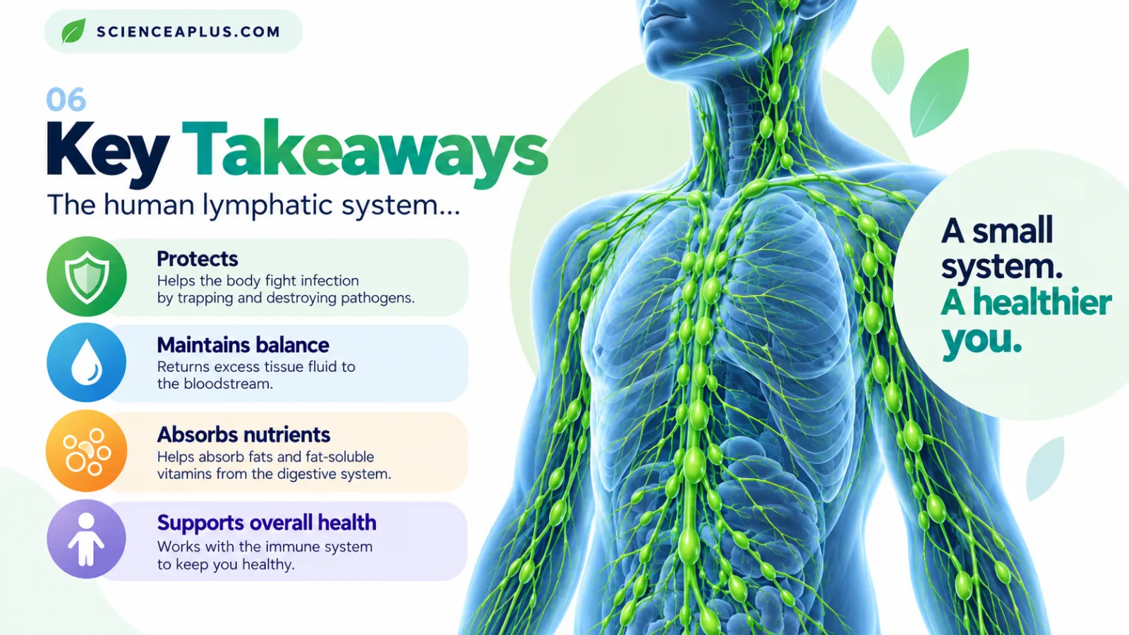
The lymphatic system is a network of vessels, lymph nodes and lymphoid organs such as the spleen, thymus and tonsils. It returns excess fluid from the tissues to the blood, absorbs fats from the gut, and helps defend the body against infection by housing and training white blood cells called lymphocytes.
What is lymph?
Lymph is tissue fluid after it has entered the lymph capillaries. It is clear to pale yellow and contains water, ions, some proteins and lymphocytes. After a fatty meal, lymph from the intestine contains fats and looks milky, and it is then called chyle.
What is the difference between lymph and blood?
Blood is pumped by the heart through closed vessels and contains red blood cells, platelets and many plasma proteins. Lymph moves in its own vessels, has little protein and no red cells, but contains lymphocytes. It is moved by muscles and valves, not by a pump.
Does the lymphatic system have a pump?
Not a central pump like the heart. Lymph is moved by skeletal muscles squeezing the vessels, pressure changes in the chest while you breathe, and contraction of smooth muscle in the walls of larger lymph vessels. Valves keep the lymph flowing one way. Being inactive slows the flow.
Where are the lymph nodes in the body?
There are several hundred lymph nodes, with estimates ranging from about 450 to 800. They occur in clusters in the neck (cervical), armpits (axillary), groin (inguinal), chest and abdomen. Each is a small bean-shaped structure, typically up to about 1–2 cm long.
Where does lymph drain into the blood?
Lymph drains into veins near the base of the neck. The thoracic duct carries lymph from the legs, abdomen, left arm and left side of the head, neck and chest into the left venous angle. The right lymphatic duct drains the right arm and right side of the head, neck and chest.
What does the spleen do?
The spleen sits in the upper left abdomen under the ribs. It filters blood, removes old or damaged red blood cells, stores some platelets and white cells, and contains lymphocytes that respond to pathogens in the blood. People can live without one but have a higher infection risk, so the NHS advises vaccines and sometimes antibiotics.
Why do lymph nodes swell?
During an infection, lymphocytes in the nodes multiply, so the nodes swell. Swollen neck nodes during a sore throat are a common example. They usually go down within 1 to 2 weeks. The NHS advises seeing a GP if they are getting bigger or have not gone down within 1 week, feel hard, or come with night sweats or a very high temperature.
How can I keep my lymphatic system healthy?
Regular physical activity, staying hydrated, keeping to a healthy weight and not smoking all support general health, and movement helps lymph flow. People with lymphoedema should follow their care team’s skin-care advice. Lymphatic detox products are not backed by good evidence.
What is lymphoedema?
Lymphoedema is long-term swelling, usually in an arm or leg, caused by the lymphatic system not draining properly. It can be inherited or develop later, for example after cancer treatment, infection or injury. The NHS describes compression garments, skin care, exercise and manual lymphatic drainage as ways of managing it.