Before starting the lesson, try to recall the structure of the human heart and the main vessels in our blood circulatory system.
Identify, the parts of the human heart.
Structure of the Human heart
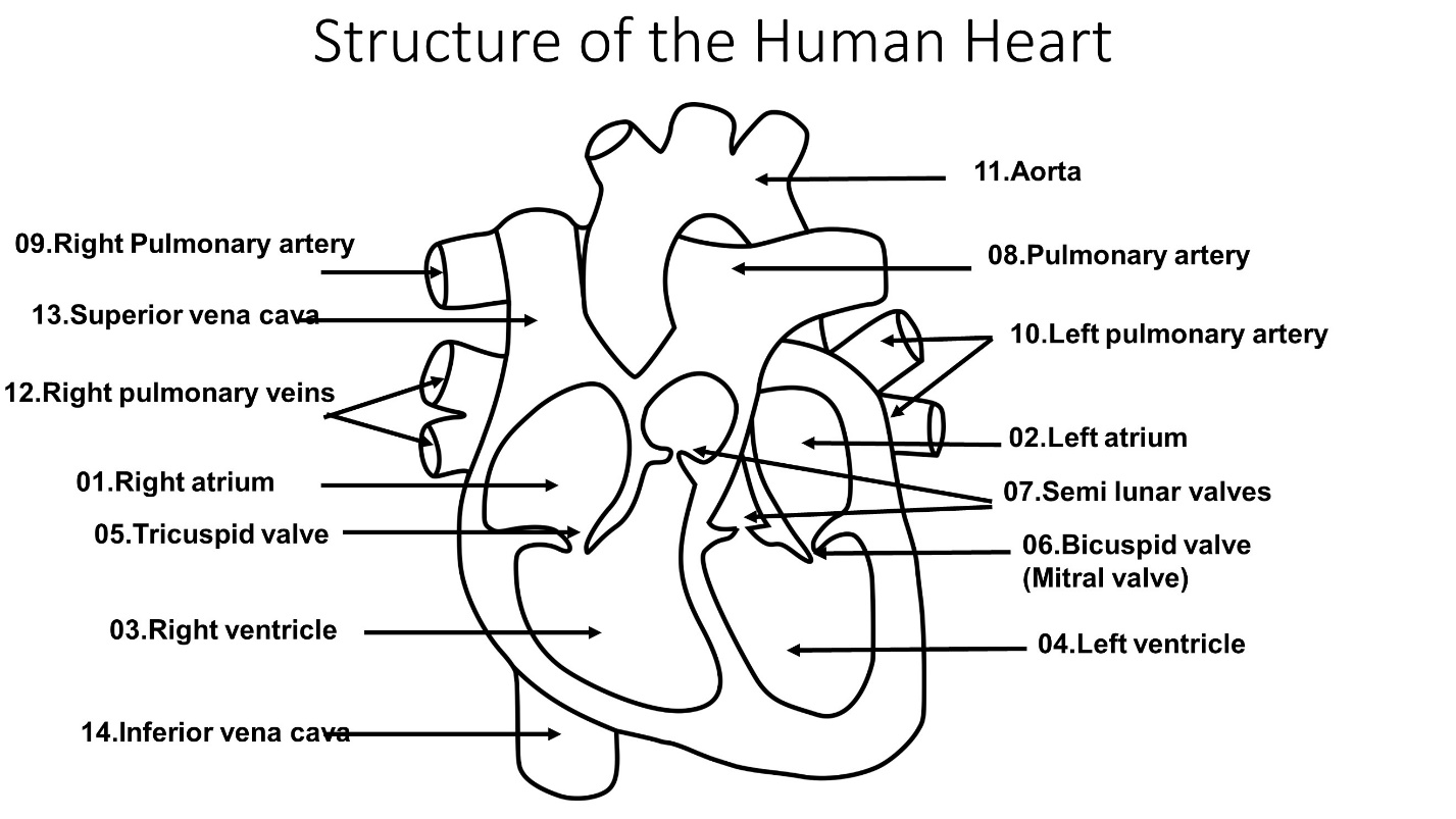
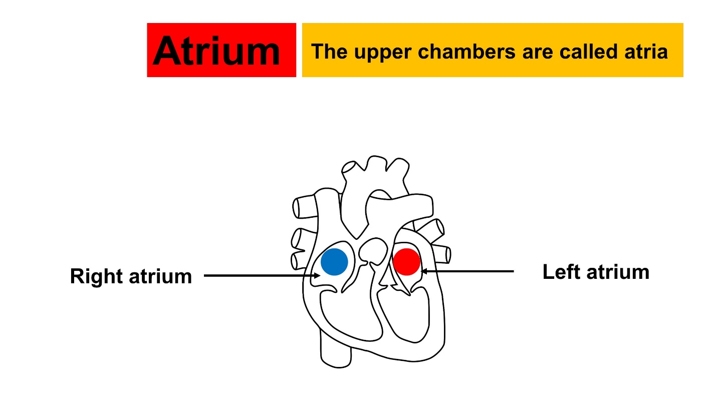
Atrium
The upper chambers are called atria.
It consists of, a Right atrium and, a Left atrium.
The left atrium contains oxygenated blood.
The right atrium contains deoxygenated blood.
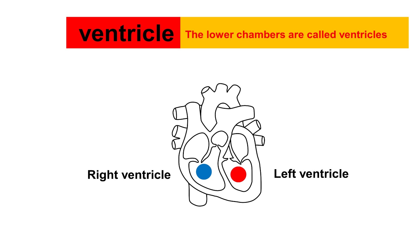
Ventricles
The lower chambers are called ventricles. Ventricles are in right and left sides.
Right ventricle pump, Deoxygenated blood to the pulmonary artery.
The left ventricle pumps oxygenated blood into the aorta.
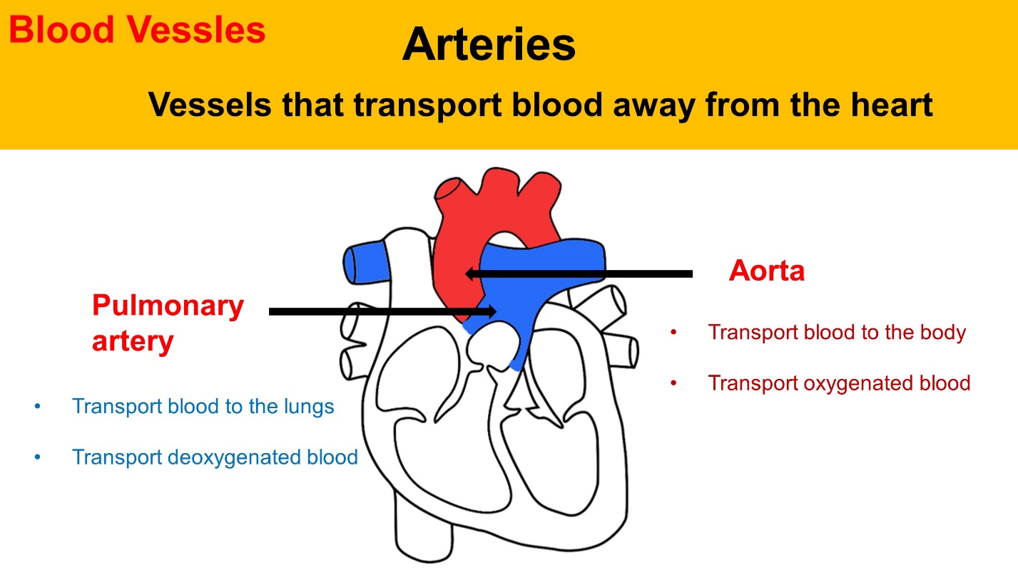
Arteries
Vessels, that carry blood away from the heart.
The main arteries are Aorta and Pulmonary artery.
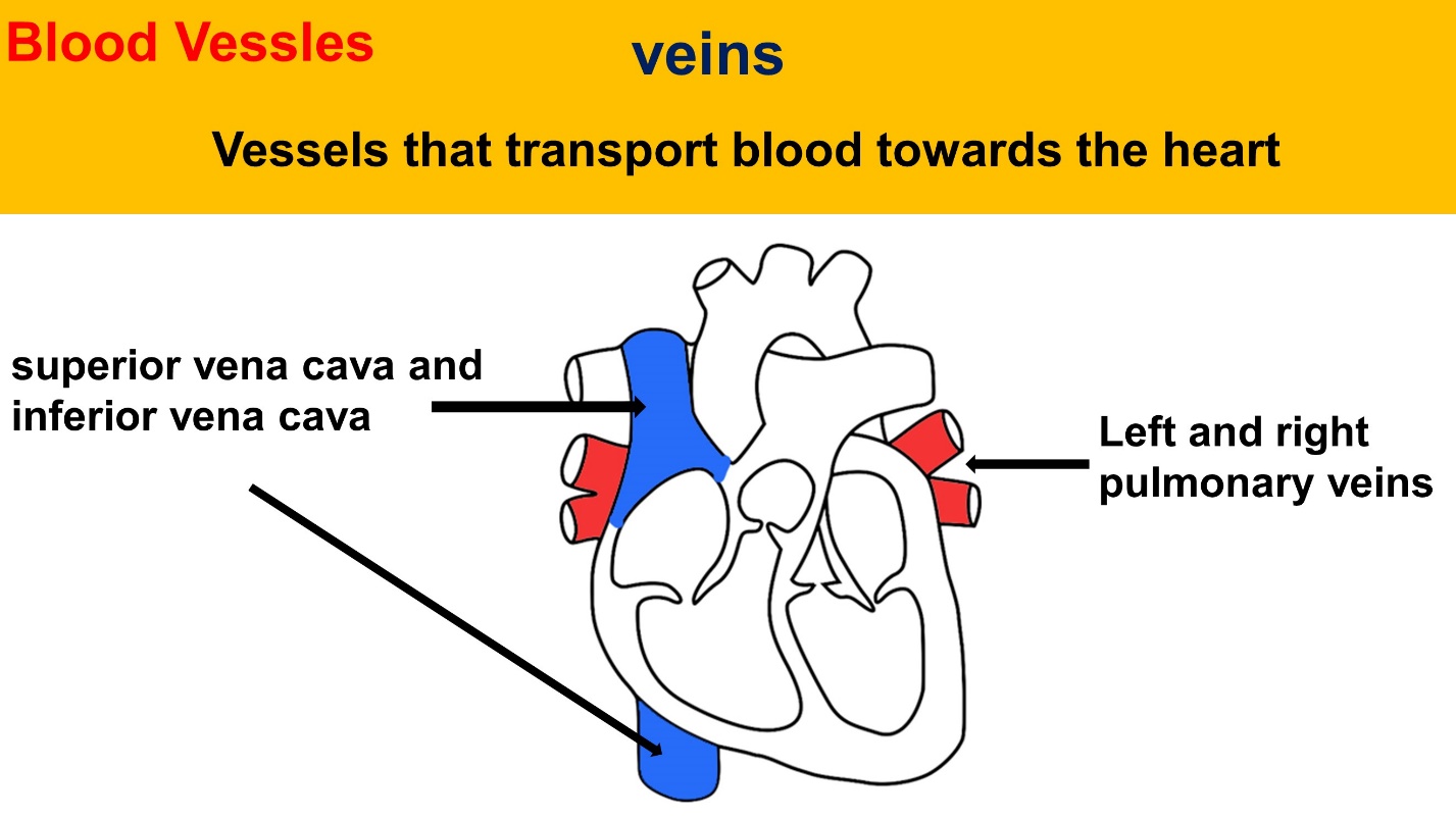
Veins
The main veins are, superior vena cava, inferior vena cava, and left and right pulmonary veins.
Cardiac muscles
The walls of the heart, are made of specific group of muscles known as cardiac muscles. Cardiac muscles can contract and then relax in cycles continuously, without any fatigue.
Cardiac cycle
The phenomenon occurs as a result of blood being transported through the heart by a sequence of contractions and relaxations of the muscles in the four-chamber walls.
As a result, the cardiac cycle is defined as a full heartbeat from its genesis through the start of the following beat.

Stages of the cardiac cycle
1. Diastole. – It happens due to atrial contraction.
2. Systole – It happens due to ventricular contraction.
3. Intervening – Atrial and Ventricular relaxation. It is called complete cardiac diastole.
- The contraction of the atria is called diastole. It takes 0.1 seconds.
- The contraction of the ventricles is called systole. It takes 0.3 seconds.
- When atria and ventricles are in relax mode, it is called, intervening. It takes 0.4 seconds.
Steps of cardiac cycle
In a summary, these are the steps of the cardiac cycle.
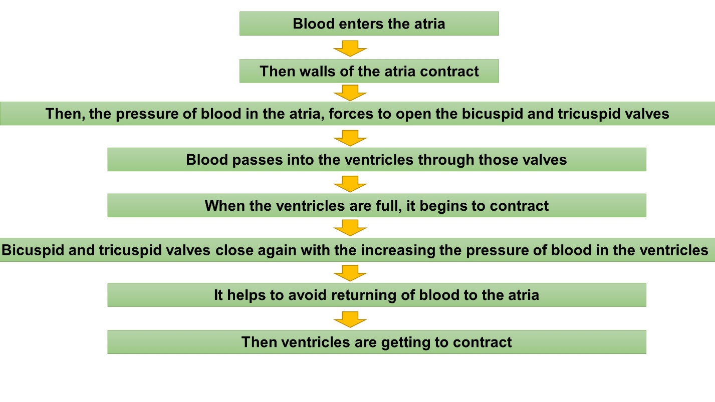
- First, Blood enters the atria.
- Then, the walls of the atria contract.
- Then, the pressure of blood in the atria, forces open the bicuspid and tricuspid valves.
- Blood passes into the ventricles, through those valves.
- When the ventricles are full, it begins to contract.
- Bicuspid and tricuspid valves close again with the increasing pressure of blood in the ventricles.
- It helps to avoid, the return of blood to the atria.
- Then ventricles contract further.
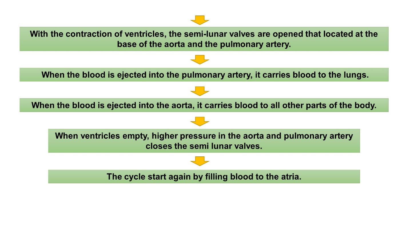
- With the contraction of the ventricles, the semi-lunar valves open.
- When the blood is ejected from the right ventricle, into the pulmonary artery, it carries blood to the lungs.
- When the blood is ejected from the left ventricle, into the aorta, it carries blood to all other parts of the body.
- When ventricles empty, higher pressure in the aorta and pulmonary artery closes the semi-lunar valves.
- The cycle starts again by filling blood into the atria.
Lub – Dup sound in the heartbeat
Lub sound is produced, when bicuspid and tricuspid valves close in atrial contraction.
- The Lub sound is longer than Dup sound.
Dup sound is results when semi-lunar valves close.
- Dup sound is shorter.
Human heartbeat
contractions and dilations of the heart muscle results in the heartbeat.
- The rate of the heartbeat of a healthy person at rest is 72 beats per minute.
Heart rate can change, according to the needs of the body
- When we exercise, the heart rate increases to supply more oxygen to the muscles.
- When we are angry or afraid, our heart rate increases. This helps to fight or run away and is called the ‘fight or flight response.
- When sleep, our heart rate decreases to provide less energy and less oxygen.
The heart rate is controlled, by nerve impulses from the medulla oblongata in the brain.
What is an electro Cardio Gram? – ECG
ECG represents the potential changes that take place in cardiac muscle cells during heart function.
This chart shows, the electrocardiogram of a healthy person.
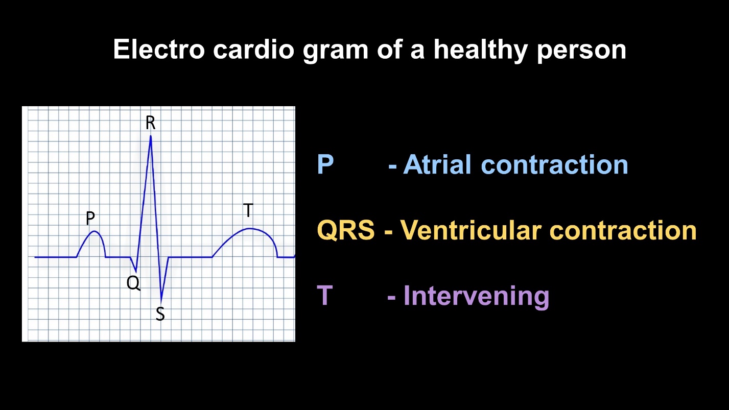
Letter P indicates, the Atrial contraction stage of the cardiac cycle.
The shape of the graph by the letters QRS indicates the ventricular contraction.
Letter T shows, Intervening.
These patterns deviate, from normal patterns due to, dysfunction of the heart.
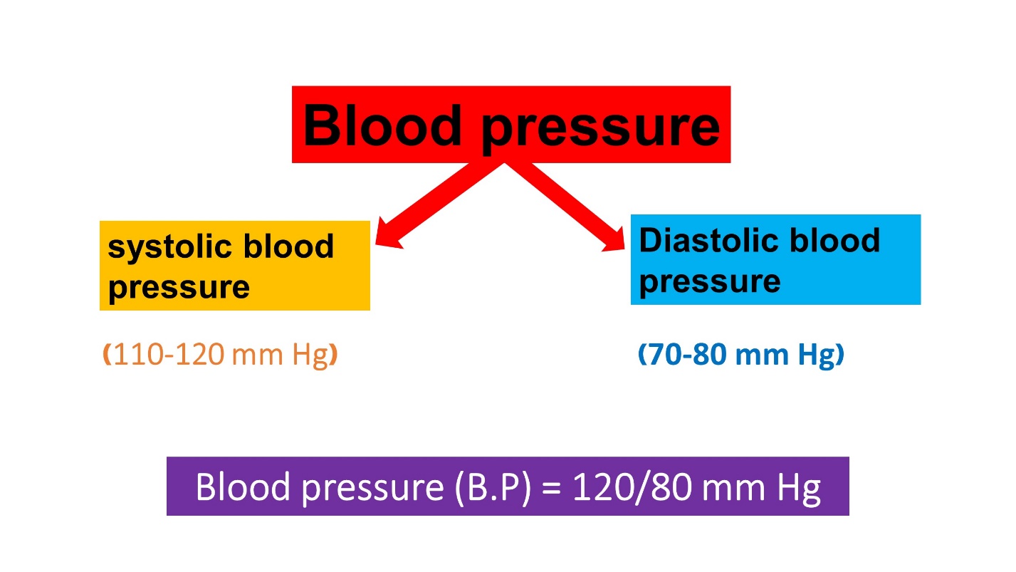
Blood pressure
The force that creates pressure on the arteries is called blood pressure.
Our blood pressure is measured as two parameters
They are,
- Systolic blood pressure
- Diastolic blood pressure
When the heart contracts and pushes blood through the arteries to the rest of the body, it creates pressure on the arteries. It is called systolic blood pressure.
The pressure in the arteries, when the heart rests between beats is called diastolic blood pressure.
- Normal diastolic blood pressure is between 70 to 80 millimeters of mercury.
This is the way of representing the value of our blood pressure.