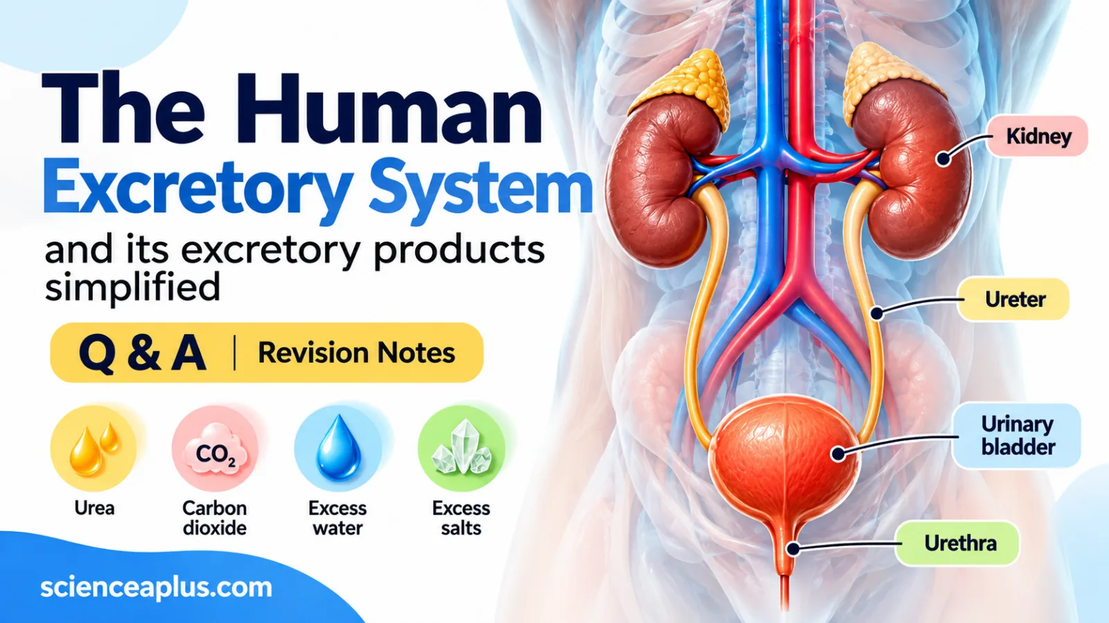
Excretion is the removal of waste products of metabolism, and of substances in excess, from the body. Every cell in your body is a tiny chemical factory, and chemical factories make waste. The human excretory system is the set of organs that clears that waste away before it can build up and cause harm.
This guide covers the human excretory system for GCSE biology, with A-level stretch boxes for anyone who wants to go further. We start with the basics: what excretion means, what the main excretory products are, and where each one comes from. Later sections look at the excretory organs, the kidney and how urine is made.
What is excretion?
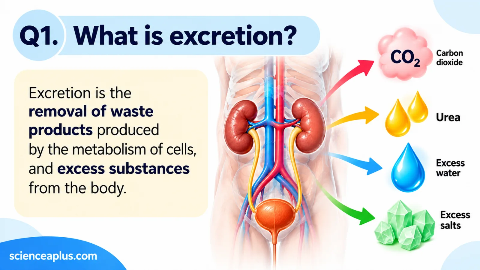
Excretion is one of the seven life processes. Living things carry out thousands of chemical reactions in their cells, together called metabolism. Some of these reactions produce substances the cell cannot use, and some leave substances in excess. Getting rid of both is excretion.
The key idea for exams is where the waste comes from. Excretory products are made inside cells, or are surplus to what the body needs. That is what separates excretion from other ways of losing material from the body.
Excretion vs egestion
Students often mix these up. Egestion is the removal of undigested food as faeces. Most of that material has never been inside a cell, because it passed straight through the gut without being absorbed. So egestion is not excretion.
| Feature | Excretion | Egestion |
|---|---|---|
| What is removed | Waste products of metabolism and substances in excess | Undigested food |
| Where it was made | Inside body cells | It never entered the cells (it stayed in the gut) |
| Examples | Carbon dioxide, urea, excess water and salts | Fibre and other material that was not digested |
| Leaves as | Breath, urine, sweat | Faeces |
Why waste must be removed
Waste products are not harmless leftovers. Some are toxic, and others upset the balance of water and salts that cells depend on. Ammonia, for example, is very toxic, which is why the body converts it into the less harmful urea before removing it.
Too much carbon dioxide changes how blood behaves. Too little or too much water and salt changes how much water moves in and out of cells. Removing waste keeps the conditions inside the body steady, which is the idea behind homeostasis.
Excretory products and where they come from
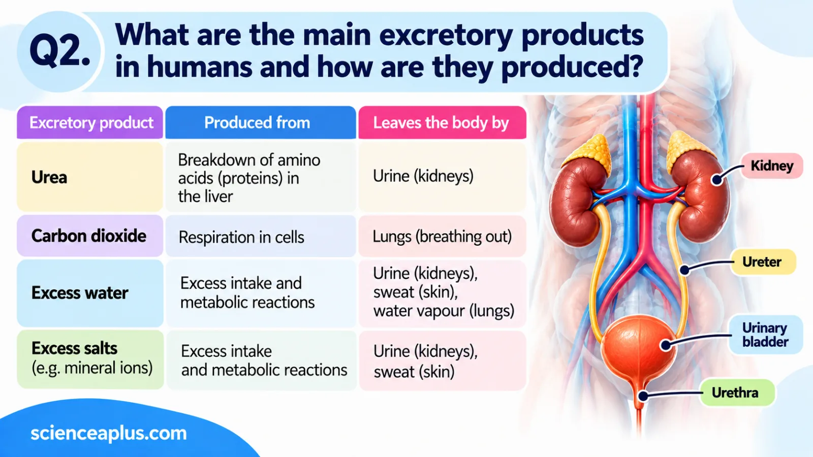
The main excretory products each come from a different process and leave by a different route. This table is worth learning, because exam questions often ask you to match a product to its source and its organ.
| Excretory product | How it is made | Removed by |
|---|---|---|
| Carbon dioxide | Aerobic respiration in cells | Lungs (in breath) |
| Urea | Liver breaks down excess amino acids (deamination) | Kidneys (in urine) |
| Excess water | Taken in as drink and food, and made in respiration | Kidneys (urine); skin (sweat); lungs (water vapour) |
| Excess ions (salts) | Absorbed from food and drink | Kidneys (urine); skin (sweat) |
| Uric acid | Breakdown of purines from DNA and RNA | Kidneys (in urine) |
| Bile pigments | Liver breaks down haemoglobin | Faeces |
Carbon dioxide
Carbon dioxide is made in every cell that carries out aerobic respiration. It diffuses out of the cells into the blood, travels to the lungs and leaves in each breath you breathe out. You can read the full story of where it comes from in our guide to cellular respiration.
Urea and deamination
Your body cannot store excess amino acids. Instead, the liver removes the amino group from each one in a process called deamination. The amino group leaves as ammonia, which is very toxic. The liver quickly converts the ammonia into urea, which is much less toxic, using the ornithine (urea) cycle.
amino acids → ammonia → urea (in the liver)
Urea then travels in the blood to the kidneys, which remove it in urine.
Excess water and salts
You take in water and salts every day in food and drink, and the amount rarely matches what you need exactly. The kidneys remove the surplus in urine. Some water and salts are also lost in sweat, and water leaves as water vapour every time you breathe out.
Because the body adjusts how much it removes, the water and salt content of the blood stays fairly constant even when you drink a lot or a little.
Uric acid and bile pigments
Two further excretory products are worth knowing. Uric acid comes from the breakdown of purines, which are found in DNA and RNA, and it leaves in urine. Bile pigments come from the breakdown of haemoglobin by the liver and leave in faeces, as we saw above.
The excretory organs
Several organs share the job of removing metabolic waste, which is why we talk about the excretory organs in the plural. Each one deals with different excretory products, and together they keep the chemistry of the body stable.
Lungs
The lungs remove carbon dioxide, the waste gas made by aerobic respiration in every living cell. Carbon dioxide diffuses from the blood into the alveoli and leaves in each breath. Water also leaves as water vapour, which you can see when you breathe onto a cold window. You can read how the airways and alveoli work in our guide to the human respiratory system.
Skin
The skin contains sweat glands. Sweat is mostly water, with some salts and a little urea. Even so, the main job of sweating is cooling the body, not excretion. Sweating does remove small amounts of waste, but it is a side effect rather than the main purpose.
Liver
The liver is closely linked to excretion because it makes two excretory products. It converts toxic ammonia, released when excess amino acids are broken down, into urea. It also makes bile pigments from the breakdown of haemoglobin. The urea travels in the blood to the kidneys, while the bile pigments leave the body in faeces.
Kidneys
The kidneys are the main organs of excretion for dissolved waste. They filter the blood, remove urea and excess water and ions, and produce urine. They also control how concentrated your blood is, which makes them central to homeostasis. The rest of this article follows the kidneys in detail, so it helps to learn this table of organs and products first.
The human urinary system
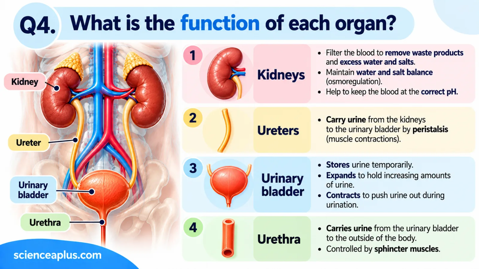
The urinary system is the part of the human excretory system that makes, carries, stores and releases urine. It has two kidneys, two ureters, one bladder and one urethra.
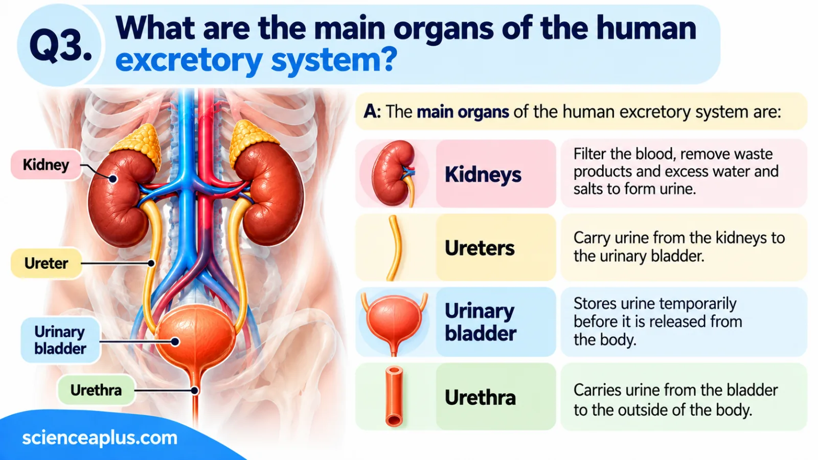
| Organ | Job |
|---|---|
| Kidneys (two) | Filter the blood and make urine; remove urea, excess water and ions |
| Ureters (two) | Carry urine from each kidney to the bladder by peristalsis |
| Bladder | Stores urine; holds roughly 300–500 mL in adults before you feel the urge to go |
| Urethra | Carries urine out of the body; controlled by sphincter muscles |
Kidney position and blood supply
The kidneys are bean-shaped and about 10–12 cm long, roughly the size of a fist. They sit either side of the spine at the back of the upper abdomen. The right kidney is slightly lower because the liver takes up space above it.
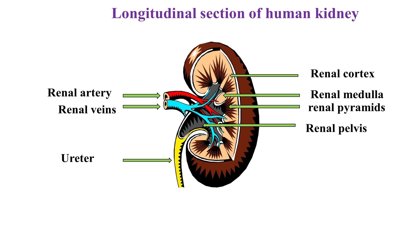
Blood arrives through the renal artery, a branch of the aorta. It leaves through the renal vein, which drains into the inferior vena cava. The kidneys receive about 20–25 % of the blood pumped by the heart, which is about 1.2 litres a minute at rest. That is a huge supply for two small organs, and it reflects how much filtering they do. To see how this fits into the whole circuit, read about double blood circulation. For more on what blood carries, see our complete lesson on human blood.
Inside a kidney
If you cut a kidney in half, you can see three main regions. The outer layer is the cortex. Inside it is the medulla, which contains triangular sections called renal pyramids. At the centre is the renal pelvis, a funnel that collects urine and passes it into the ureter.
Each kidney contains about 1 million nephrons, the tiny functional units that do the filtering. A nephron has a Bowman’s (glomerular) capsule around a knot of capillaries called the glomerulus, then a proximal convoluted tubule, a loop of Henle, a distal convoluted tubule and a collecting duct. The next section explains how these parts turn blood into urine.
How urine is formed in the nephron
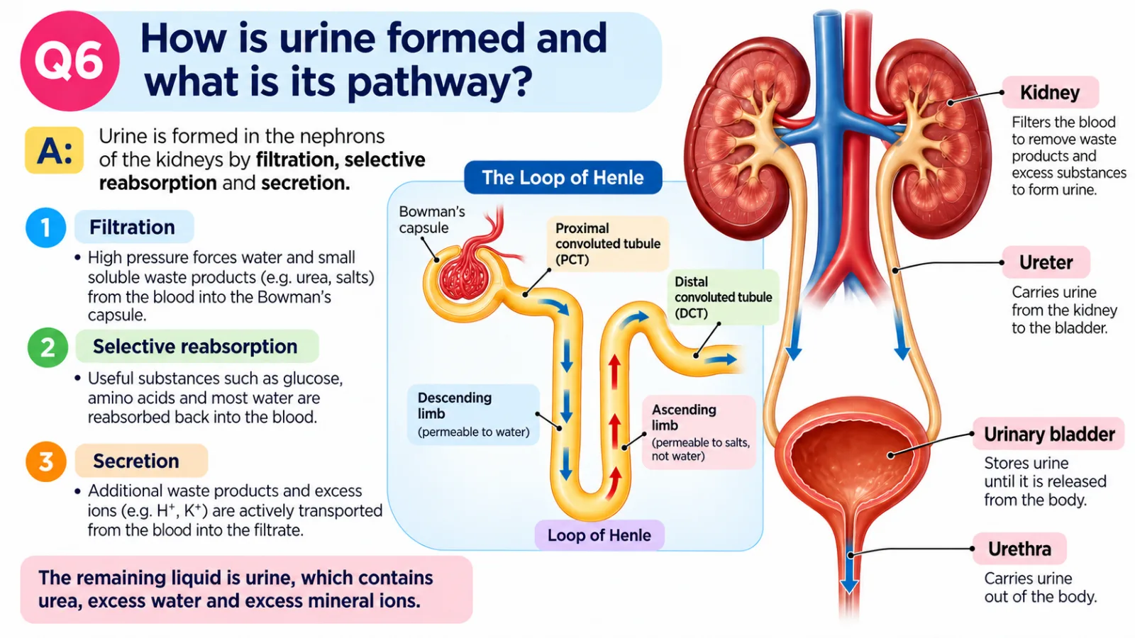
Of all the excretory organs, the kidneys do the most delicate work. Each kidney contains about 1 million nephrons, and every nephron is a tiny filtering and recycling unit. Together they turn blood into urine in a continuous, four-stage process: ultrafiltration, selective reabsorption, water control and secretion.
A nephron has five parts, in this order: the Bowman’s (glomerular) capsule wrapped around a glomerulus (a knot of capillaries), the proximal convoluted tubule, the loop of Henle, the distal convoluted tubule, which then drains into a collecting duct shared by many nephrons. Keep that sequence in mind as you read, because exam questions often ask you to say where each step happens.
Step 1: Ultrafiltration
Blood arrives at the glomerulus through a wide afferent arteriole and leaves through a narrower efferent arteriole. Because the way out is narrower than the way in, pressure builds up inside the glomerulus. This high pressure forces small molecules out of the blood and into the capsule: water, glucose, amino acids, urea and ions.
Blood cells and large proteins are too big to pass through, so they stay in the blood. The liquid that enters the capsule is called filtrate. Around 180 litres of it are made every day, which is far more than the body could ever afford to lose.
Step 2: Selective reabsorption
Most of that filtrate is useful, so the proximal tubule takes it back. Normally all of the glucose is reabsorbed here, along with amino acids and much of the water and salt. Two processes do the job. Glucose and many ions are moved back into the blood by active transport, which uses energy. Water then follows by osmosis.
Some substances can also move back by diffusion, down their concentration gradient. The key idea is that the kidney is selective: it keeps what the body needs and leaves what it does not. Only part of the urea is reabsorbed, so much of it stays in the tubule and becomes more concentrated as water is taken back.
Step 3: Controlling water
Further along, in the collecting duct, the amount of water reabsorbed is adjusted to suit the body’s needs. The loop of Henle sets up the salty medulla that makes this possible. This is where the hormone ADH steps in, which is covered in the next section.
Step 4: Secretion
Finally, some substances are actively added into the tubule from the blood. Examples include hydrogen ions (H⁺), potassium ions (K⁺) and some drugs. Secretion helps to fine-tune the composition of the blood and gets rid of extra excretory products that filtration alone might miss.
Where did all that water go?
Put the numbers side by side and the scale of reabsorption becomes clear. About 180 litres of filtrate are made each day, yet only about 1–2 litres leave the body as urine. That means roughly 99 % of the filtrate is reabsorbed.
180 litres of filtrate but only about 1.5 litres of urine: try the calculator below to see what percentage that is, and what happens when you change the numbers.
The loop of Henle (A-level stretch)
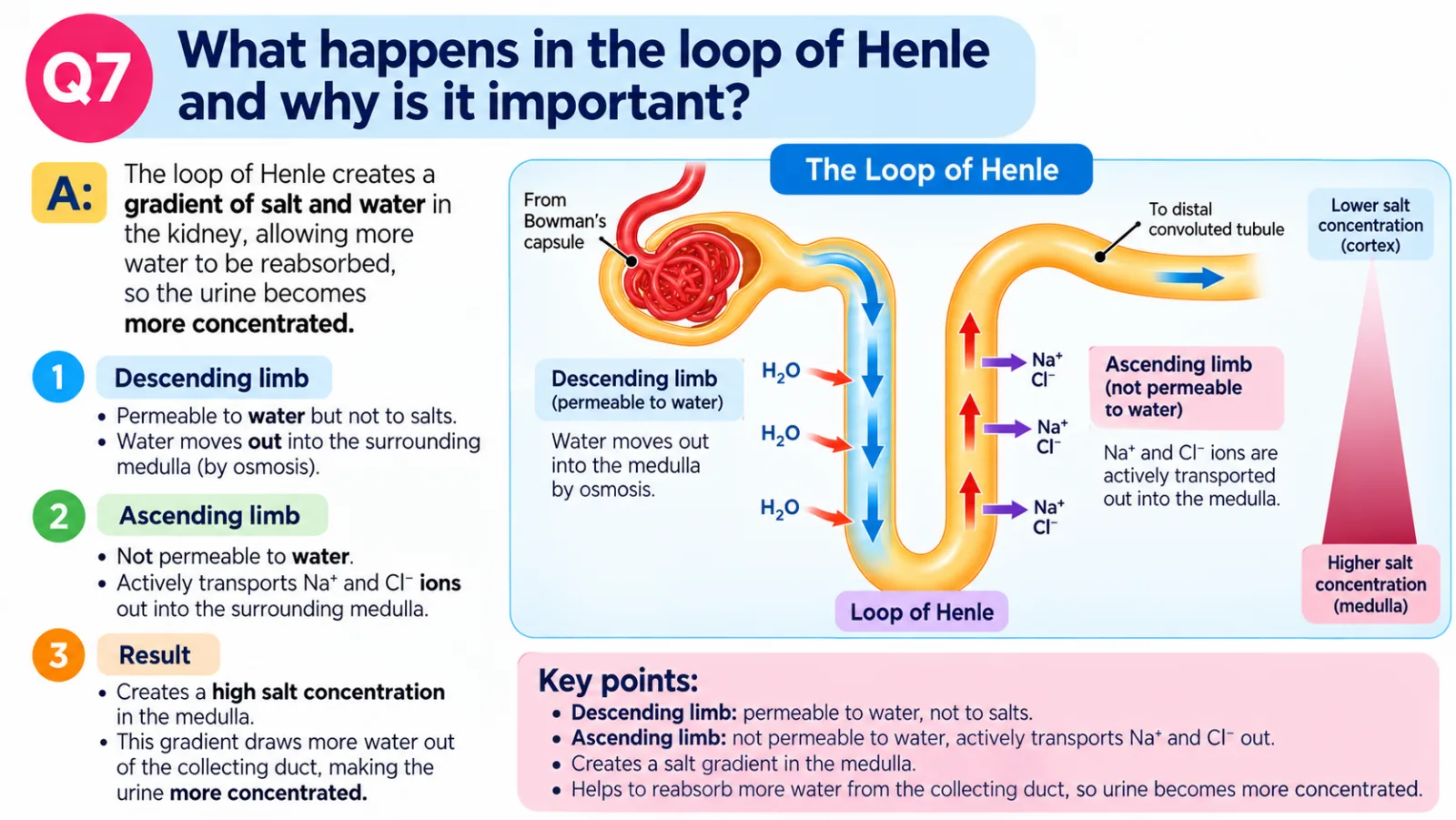
The loop of Henle dips down into the medulla and back up again. Its two limbs behave very differently. The descending limb is permeable to water. The ascending limb is impermeable to water, and it pumps sodium (Na⁺) and chloride (Cl⁻) ions out into the surrounding tissue.
This pumping creates a high salt concentration in the medulla. When the filtrate later passes down the collecting duct, water can be drawn out by osmosis into that salty tissue and returned to the blood. The more water that is drawn out, the more concentrated the urine becomes.
Controlling water: ADH and osmoregulation
The body must keep the water content of the blood within narrow limits. This is called osmoregulation, and it is an example of homeostasis. The kidneys do the adjusting, and a hormone called ADH (antidiuretic hormone) from the pituitary gland sends the instructions.
The system works by negative feedback, which means any change is detected and then reversed. Here is the loop for a person who is becoming dehydrated:
- The person sweats, breathes out water vapour or does not drink enough, so the blood becomes too concentrated.
- The brain detects the change and the pituitary gland releases more ADH into the blood.
- ADH travels to the kidneys and makes the collecting ducts more permeable to water.
- More water is reabsorbed from the filtrate back into the blood.
- The kidneys produce a small volume of concentrated urine, and the blood becomes more dilute again.
- Once the blood water level is back to normal, ADH release falls. This is the negative feedback that stops the response going too far.
If the blood is too dilute, for example after drinking a lot of water, the process runs the other way. Less ADH is released, the collecting ducts become less permeable, and a large volume of dilute urine is produced.
This links back to the bigger picture of the human excretory system. Excretion is not only about removing urea. It also covers removing excess water and ions, and ADH is how the kidneys decide how much of each to remove.
Check your understanding
Tap a question to reveal the model answer, but try to answer it in your head first.
Explain why a person who has not drunk anything all day produces a small volume of dark urine. (3 marks)
The blood becomes too concentrated (1). More ADH is released from the pituitary gland, which makes the collecting ducts more permeable to water (1). More water is reabsorbed into the blood, so a small volume of concentrated urine is produced (1).
Describe what happens in the glomerulus during ultrafiltration, and name two substances that are not filtered out. (4 marks)
The afferent arteriole is wider than the efferent arteriole, so high pressure builds up in the glomerulus (1). This forces small molecules such as water, glucose, amino acids, urea and ions out of the blood into the Bowman’s capsule (1). Blood cells (1) and large proteins (1) are too big, so they stay in the blood.
What is in urine?
Urine is the main route by which the human excretory system gets rid of dissolved wastes. It is about 95 % water, with urea, salts and other waste substances dissolved in it. The exact mixture changes from hour to hour, because the kidneys adjust it to keep the blood balanced.
What is not in healthy urine matters just as much. Because of selective reabsorption, normal urine contains no glucose and no protein. If either appears, doctors treat it as a clue worth following up.
| Substance | In normal urine | Possible warning sign (doctors test for this) |
|---|---|---|
| Water | About 95 % of the volume | Volume and concentration vary with how much you drink and sweat |
| Urea | Present, from the breakdown of excess amino acids | Not used as a simple home check |
| Salts (ions) | Present, in amounts that depend on diet and sweating | Tested in blood and urine by professionals |
| Glucose | Normally absent | Glucose in urine can be a sign of diabetes |
| Protein | Normally absent | Protein in urine can indicate kidney damage |
This table is for revision only. It cannot tell you anything about your own health. If you are worried about changes in your urine or how you feel, speak to a GP or call NHS 111.
When kidneys fail: dialysis and transplants
If the kidneys stop working properly, urea and excess water build up in the blood. The two main treatments are dialysis and a kidney transplant.
Haemodialysis
In haemodialysis, the patient’s blood passes over a partially permeable membrane, with dialysis fluid on the other side. Small molecules such as urea and excess ions diffuse out of the blood into the fluid. Blood cells and large proteins stay in the blood.
The fluid is made with the same glucose and useful ion concentrations as normal blood, so those substances are not lost. It contains no urea, so urea keeps diffusing out down its concentration gradient. A patient usually needs about 3 sessions a week, each lasting about 4 hours.
Kidney transplant
A transplant replaces the failed kidney with a healthy one from a donor. It can free a patient from regular dialysis, but there is a risk of rejection, where the immune system attacks the new organ. Patients therefore need to take immunosuppressant drugs.
Trace the journey of a urea molecule
Here is the whole story of one urea molecule, from the liver to the toilet. Use it to link the liver, blood and kidneys together in your head.
- You eat more protein than your cells need, so some amino acids are in excess. The body cannot store them.
- In the liver, enzymes carry out deamination. The amino group is removed from each amino acid and released as ammonia, which is very toxic.
- The ammonia is converted into urea, which is less toxic, in the ornithine (urea) cycle.
- Urea dissolves in the blood plasma and is carried around the body until the blood reaches a kidney through the renal artery.
- In the glomerulus, high pressure forces small molecules, including urea, out of the blood and into the Bowman’s capsule as filtrate.
- The filtrate flows along the tubule and collecting duct. Useful substances such as glucose are reabsorbed, and urea leaves the kidney in the urine, collecting in the renal pelvis.
- Urine moves down the ureter by peristalsis into the bladder, is stored, and then leaves the body through the urethra when the sphincter muscles relax.
Test yourself
Key points to remember
Frequently asked questions about the human excretory system
What is excretion in biology?
Excretion is the removal of waste products of metabolism, which are made inside cells, and of substances in excess. Examples include carbon dioxide from respiration, urea from the breakdown of amino acids, and surplus water and salts. It keeps the body’s internal conditions stable.
What is the difference between excretion and egestion?
Excretion removes waste made by cells, such as urea and carbon dioxide. Egestion removes undigested food as faeces, most of which never entered cells. One nuance is that bile pigments, made by the liver from broken-down haemoglobin, are excretory products even though they leave in faeces.
What are the main excretory organs?
The main excretory organs are the lungs, kidneys and skin. The liver is closely involved because it makes urea and bile pigments. The lungs remove carbon dioxide and water vapour. The kidneys remove urea, excess water and salts in urine. The skin loses sweat. The liver makes urea and bile pigments, processing wastes for removal.
How is urea made in the body?
Urea is made in the liver. Excess amino acids undergo deamination, where the amino group is removed as ammonia, which is very toxic. The ammonia is then converted into less toxic urea in the ornithine (urea) cycle. The blood carries urea to the kidneys for removal.
What is the function of the kidney?
The kidneys filter the blood and make urine. They remove urea and excess ions and control how much water the body keeps, using the hormone ADH. Each kidney has about 1 million nephrons, the tiny filtering units where filtration, reabsorption and secretion take place.
How is urine formed?
Urine forms in the nephron in stages. High pressure in the glomerulus forces small molecules into the capsule (ultrafiltration). Useful substances such as all glucose are reabsorbed. The loop of Henle builds a salty medulla, water reabsorption is then controlled in the collecting duct by ADH, and some substances are secreted into the tubule.
What is in urine?
Urine is about 95 % water, with urea, salts and other waste substances dissolved in it. Healthy urine normally has no glucose or protein. Glucose can be a sign of diabetes and protein can indicate kidney damage, so doctors test for them. Anyone worried should see a GP.
Is sweating a form of excretion?
Partly. Sweating’s main job is cooling the body, but sweat also contains water, salts and a little urea, so some excretion does happen through the skin. The kidneys remain the main route for removing urea and excess salts from the body.
How does dialysis work?
In haemodialysis, blood passes over a partially permeable membrane with dialysis fluid on the other side. Urea and excess ions diffuse out into the fluid, while blood cells and proteins stay in. The fluid has normal blood levels of glucose and useful ions, and no urea. Sessions are usually about 3 a week.