Introduction
The lymphatic system in human beings is a specialized network of vessels that run throughout the body. Just like arteries and veins flow blood in the human body, lymphatic vessels flow a special fluid called lymph. Lymph has its own functions in the human body. Lymphatic vessels ensure the optimum flow of lymph. This lesson is a simplified version of the human lymphatic system, its basic anatomy, and its physiology.
Anatomy
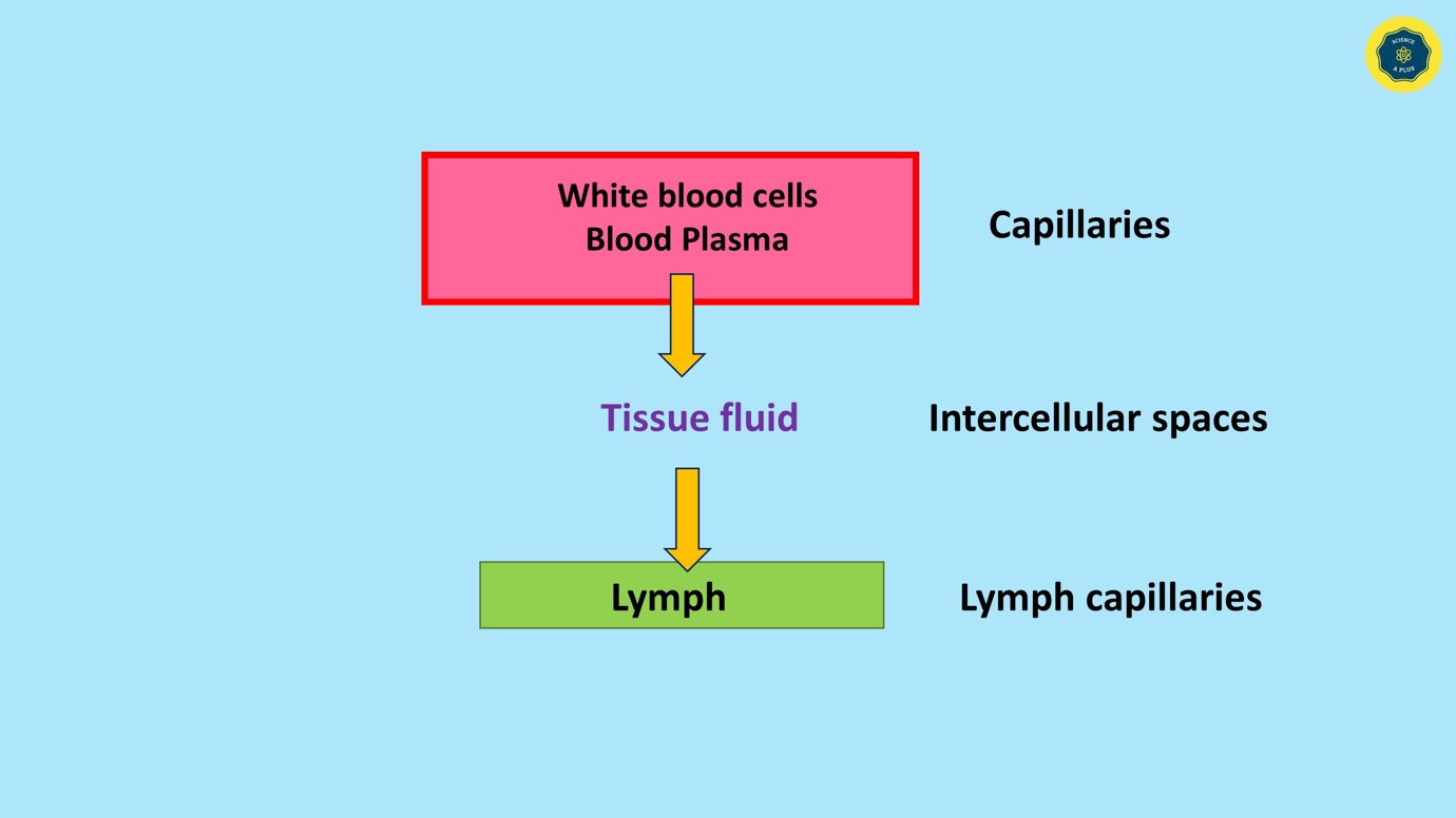
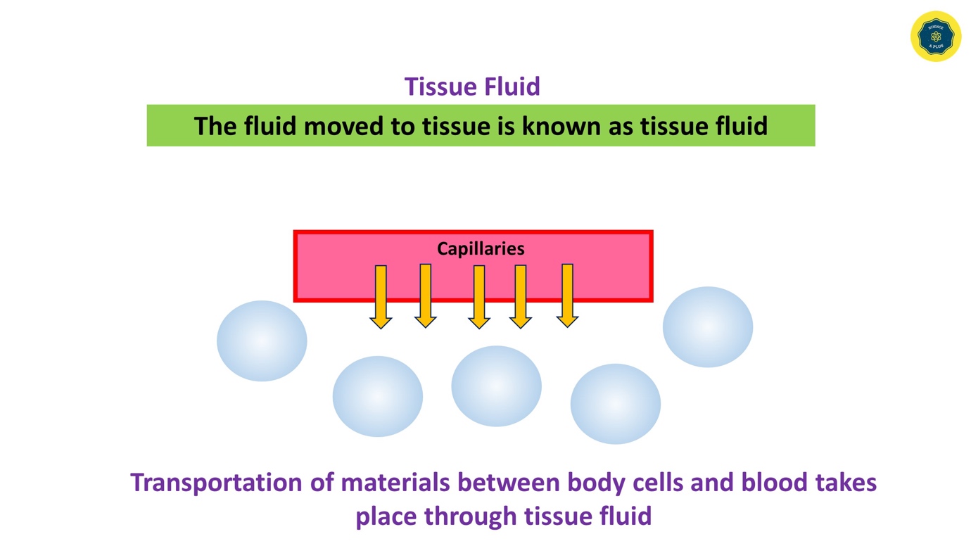
Blood plasma at the capillary level forms tissue fluid. This is formed when plasma at the capillary level enters into tissue spaces. This is discussed in detail in the next paragraphs.
When tissue fluid enters into the lymphatic system it is known as the Lymph. Lymph flows in lymphatic vessels / lymphatic ducts.
The lymphatic system consists of;
- Lacteals
- Lymphatic capillaries
- Lymph nodes
The flow of lymph in the lymphatic system is supported by the power of adjacent muscle contraction. Parts of the lymphatic system are described further in the next paragraphs.
Structure of Lymphatic ducts
Lymph flows in lymphatic ducts are there are located all over the body. To understand it simply lymph vessels or lymphatic ducts are found in all four limbs, chest abdomen, and head along vessels but as a separate structure.
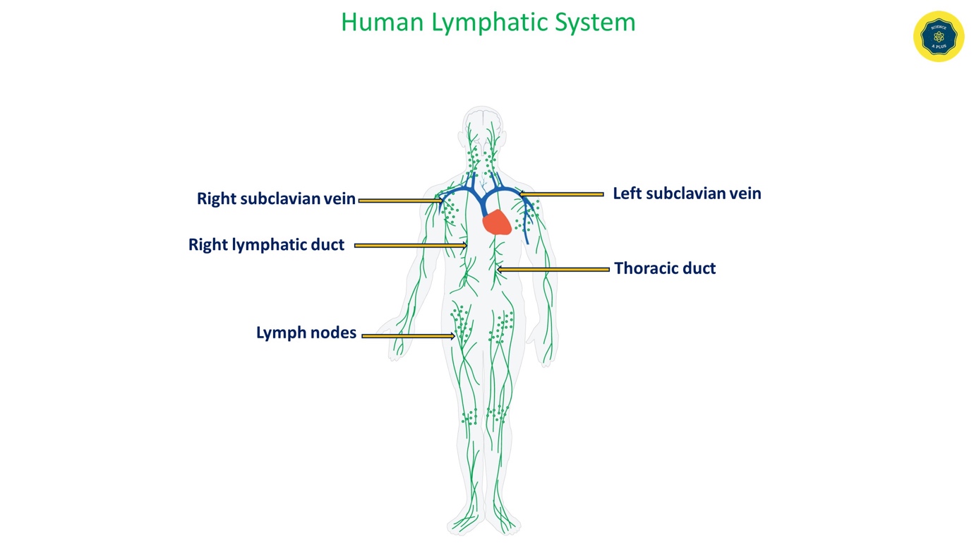
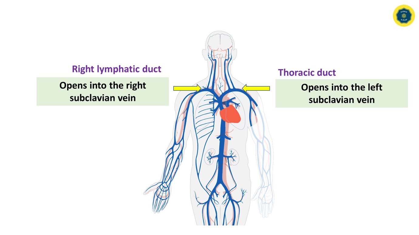
The small vessels open into two main lymphatic ducts in the human body. They are the right lymphatic duct and the thoracic duct which is found on the left side of the body. The following diagram shows the location of these vessels.
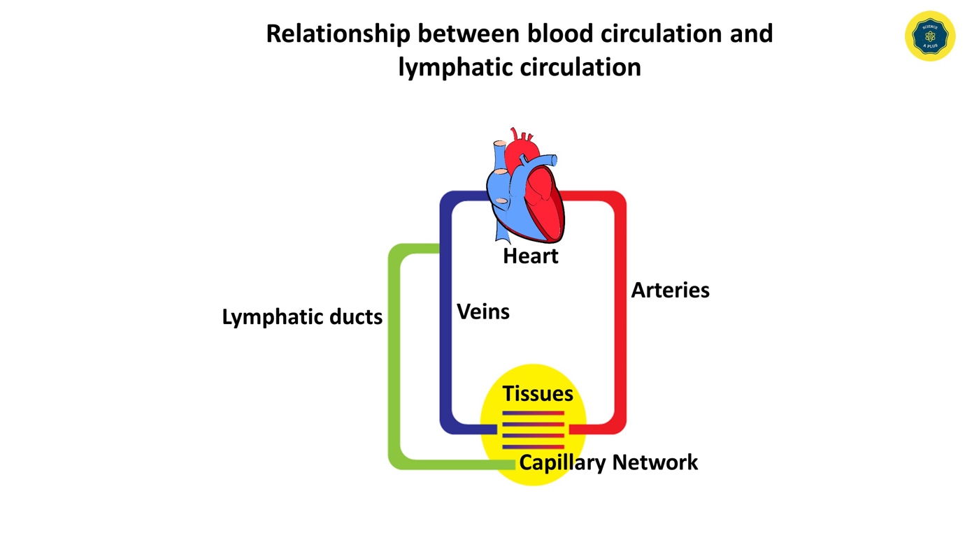
After doing the specified functions lymph flows back into blood circulation. This is aided by the above-mentioned two major lymphatic ducts. (Right lymphatic duct and the thoracic duct)
The right lymphatic duct opens into the right subclavian vein of the circulatory system. The right subclavian vein flows blood from the right hand and right side of the head to the heart.
The other main lymphatic duct, the thoracic duct opens into the left subclavian vein. The left subclavian vein flows blood from the left side of the head and left hand.
Structure of Lacteals
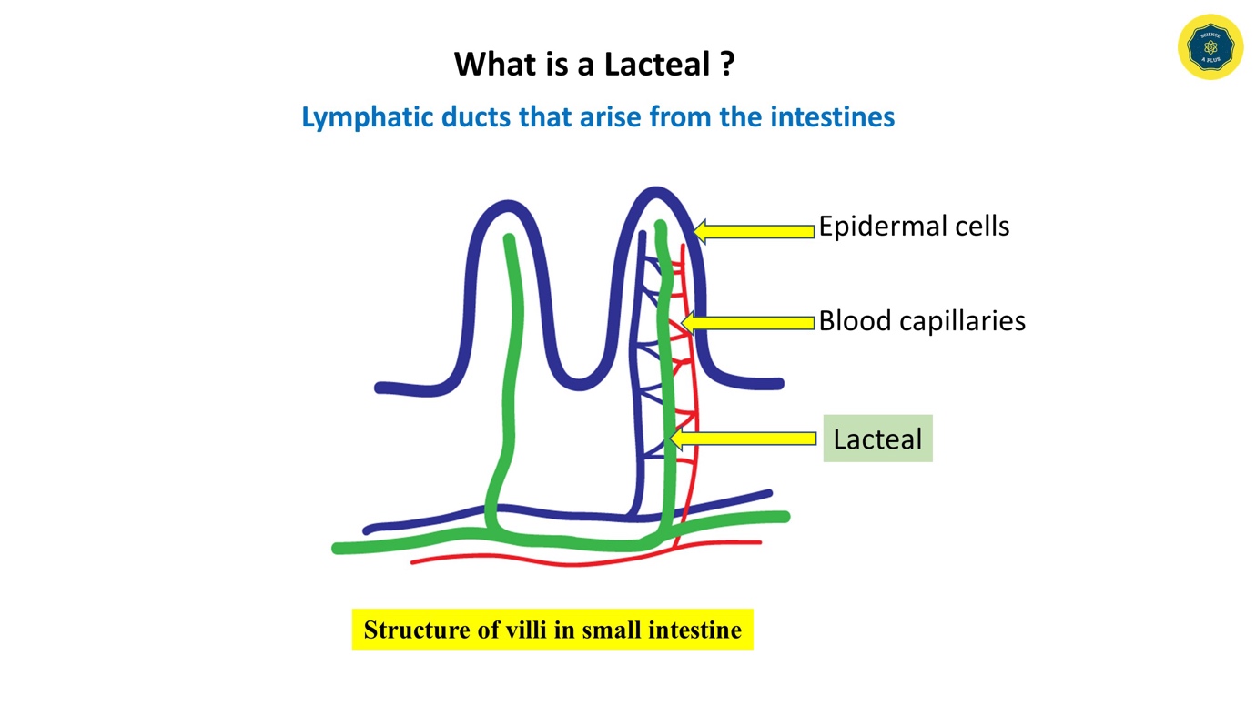
Lacteals are the lymphatic vessels that arise from the intestinal villi. They are very small and contains nutrients from food.
Lymph Nodes
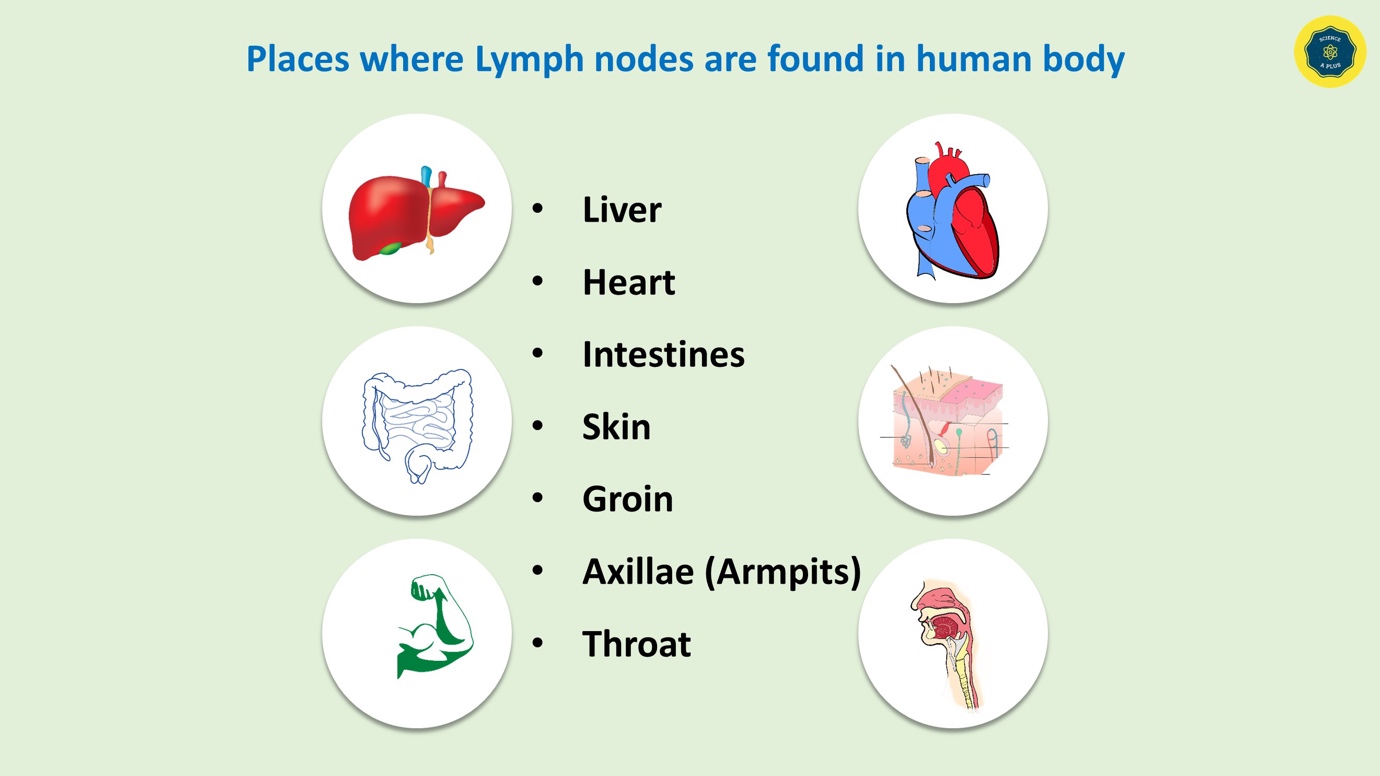
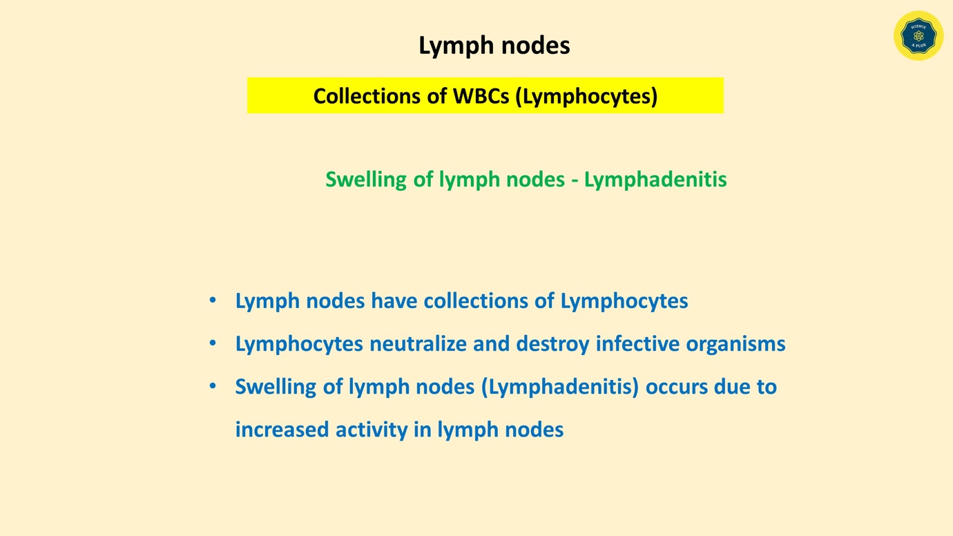
Lymph nodes are anatomical structures made with collections of WBCs. Lymph nodes are found around the liver, heart, intestines, skin, groin and axillae, and throat.
When lymph nodes are swollen it is called lymphadenitis. This is a common phenomenon in viral fevers. It happens because lymphocytes (type of WBCs) have increased activity during times of infections to protect the body by neutralizing the infective organisms.
Formation and circulation of tissue fluid
Blood flowing from the heart through the aorta reaches the end organs and cells through arteries and then arterioles and capillaries. In other words, oxygenated blood from the left side of the heart enters into the aorta and thereafter blood flows in arteries end up in capillaries where diffusion of nutrients and oxygen to tissues takes place.
At this capillary level, some of the blood plasma moves into the tissue spaces. This fluid is called the tissue fluid. Tissue fluid aids in the transportation of materials.
Tissue fluid can contain white blood cells also known as WBCs and plasma of the blood. Usually, red blood cells or else RBCs are not found in the tissue fluid. Only some types of plasma proteins are found in the tissue fluid.
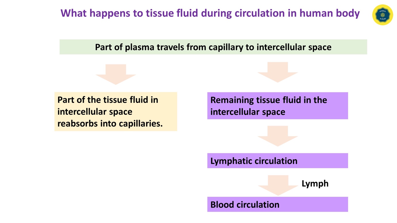
During the circulation of tissue fluid part of it is reabsorbed into capillaries whereas the remainder in the intercellular space enter into the lymphatic circulation and forms the lymph. Lymphatic circulation with its lymph finally opens into blood circulation.
Functions of Lymph
The main function of the lymph is to protect the human body from disease-causing organisms. The lymphatic system can be considered as a part of the immune system as well.
Other than that body fluid level regulation is done by the lymph to a certain extent. Lymphatics, especially lacteals aid in digestion as well.
Lymph also has a function related to the transportation of waste material from the cellular level.
