Introduction
MRI is a technique used in medicine to visualize the inner structures of the body for diagnosing diseases. This is different from X-rays, CT scans, and PET scans. MRI does not use ionizing radiation which can be harmful to the human body. MRI uses strong magnetic fields as well as radio waves to create images of the inside of the human body.
MRI scans take longer periods of time and usually louder. The patient has to lie and stay in a narrow tube in the MRI machine until the images are generated. MRI is an expensive solution and widely used in the healthcare setting with the purpose of diagnosis and staging diseases. MRI scans are usually done on a selected area of the body. That means the common MRI scans include MRI of brains, heart, gastrointestinal tract, spinal cord, knee joint, etc.
How the MRI machine works
The human body consists of tissues that mostly contain water (H2O).
Hydrogen ions / Protons are found in all tissues. These protons can be provided energy by a strong magnet. MRI scanner has strong rotating magnets that provide a strong magnetic field. This energy is absorbed into the protons of all tissues.
When the magnetic field is removed, the protons start releasing the absorbed energy. In this process, the protons emit radio-frequency (RF) waves. These RF waves can be identified and recorded by the coils in the MRI scanner. Various tissues and structures emit RF waves in different patterns and different densities.
Computers connected to the MRI scanner can control the movements of the magnet and create the image from the receiving signals. The final result is a series of images in desired planes that visualizes the inner structures of the human body. MRI is time-consuming as well as expensive.
Uses of MRI
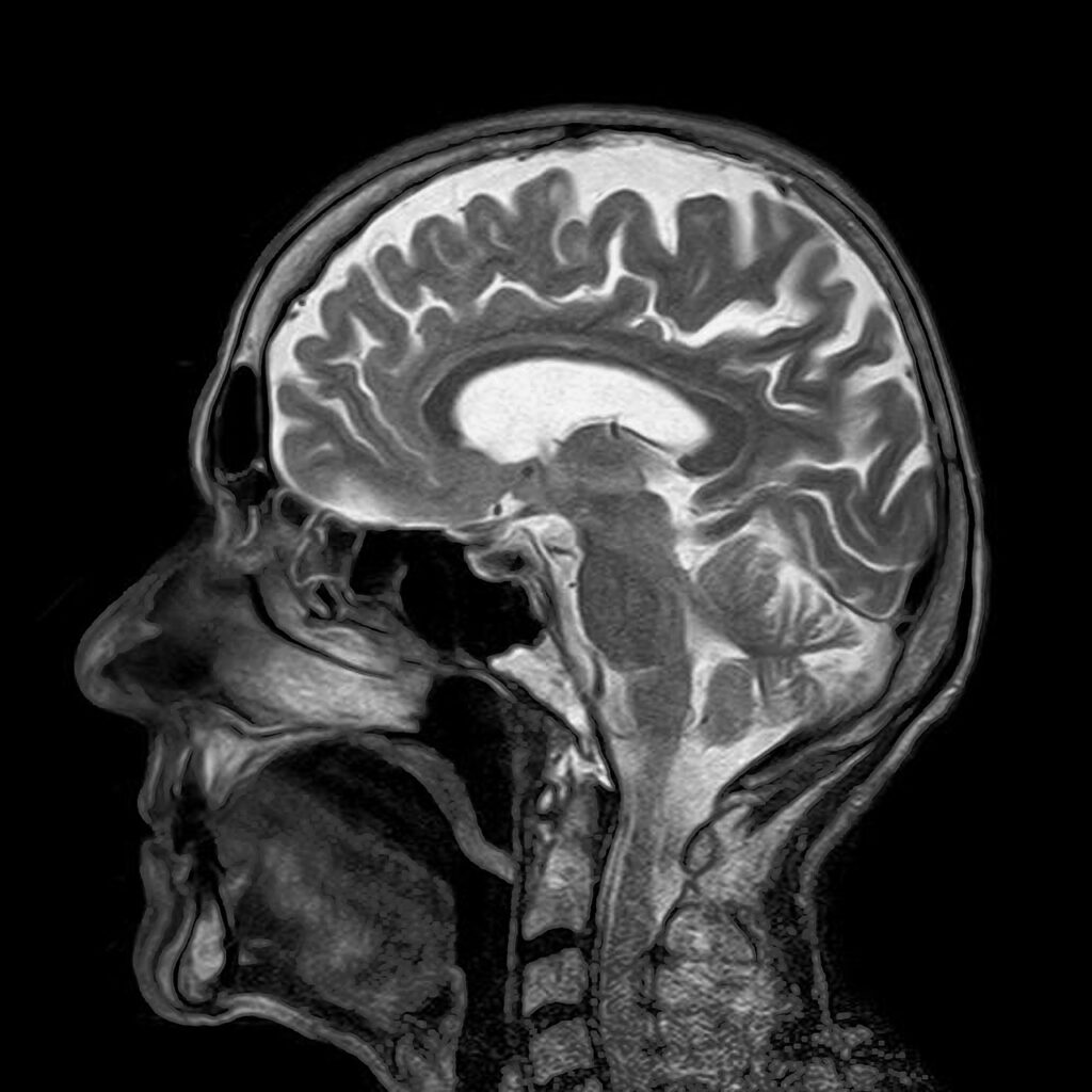
MRI is used in both medical as well as non-medical fields. The following image is taken from MRI brain.

MRI scans of the brain are used in stroke, cerebrovascular disorders, brain infections, dementia, brain tumors, and nerve degenerating diseases, and many more.
MRI of the heart can be used for identifying congenital heart diseases, inflammatory conditions of the heart, anatomical defects and functional defects of the heart, vascular disorders of the heart, etc.
MRI scans focusing on the gastrointestinal tract can be used to visualize the gallbladder, liver, pancreas, etc.
Furthermore, MRA (Magnetic Resonance Angiography) is another technique used to visualize the blood vessels of the human body. This enables the identification of the abnormal constriction (stenosis) of blood vessels as well as the abnormal dilatation of blood vessels (aneurysms). In MRA a contrast material is injected into the blood to properly visualize the structures. Gadolinium is used as the contrast material in these studies.
MRI is also used in MRI-guided surgeries to localize lesions in the body during the surgery.
MRI is generally known as safe compared to CT because MRI does not use ionizing radiation.
Other than in the healthcare setting, the MRI technique is also used in other industries. In those fields, MRI is used for the analysis of chemicals and molecular structures. Corrosion of metals is also studied with different MRI techniques.


